Digital Holographic Microscopy
OPUS V2
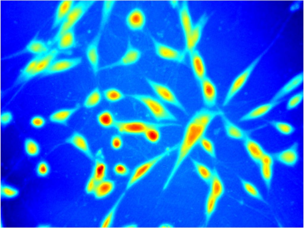
Quantitative Phase Microscopy (QPM) stands out among a suite of modern label-free imaging techniques as extremely capable high-contrast approach. Using sample’s refractive index as intrinsic contrast agent QPM numerically converts, upon phase reconstruction, recorded interference pattern into a nanoscale-precise subcellular-specific 2D/3D/4D maps of examined transparent specimen. This non-phototoxic imaging technique brings biology and metrology closer as it generates quantitative map of live bio-structure (cell mass, volume, surface area, and their evolution in time) upgrading visualization of the sample to its stain-free non-invasive optical measurement enabling precise diagnostics. Majority of QPM solutions are full-field optical techniques relying on the virtue of optical complex amplitude retrieval from recorded intensity pattern – interferograms – generated upon interference of object and reference beams. Extremely important step of each full-field fringe-based QPM method comprises interferogram phase demodulation understood as optical phase delay map decoding from the recorded intensity distributions. The need for deploying a reconstruction algorithm directly influences available information space-bandwidth-product (SBP) of the system determining quality and resolution of the measurement/imaging.


In the PHAICELL project we aim at highlighting the problem of losing information upon optonumerical processing (interferogram acquisition and phase reconstruction) fundamentally limited by noise and phase retrieval imperfections. Although novel numerical techniques based on, e.g., variational/empirical decomposition and further Hilbert spiral transform demodulation, showcase significant advancements, i.e., single-frame analysis with partially-overlapping spectrum, we are claiming the strong need for taking a step further in their development. In the PHAICELL project we unprecedentedly aim at exploring and defining the limits of QPM in terms of high-speed imaging in photon starving regime, extremely low signal to noise ratio of interferograms and strong object absorption, and push these limits. Innovative numerical framework will be based on the paradigm of image decomposition through Adaptive Local Iterative Filtering (ALIF) via originally tailored novel solutions (i.e., 2D fast ALIF). Thus, a new class of phase reconstruction techniques will be established, advancing QPM capacity. PHAICELL has unique features in terms of international network of excellence-driven partner institutions and very ambitious scientific novelty-driven plan. As a result, we plan to enable unique applications in biomedical imaging through numerically and experimentally increased SBP, especially taking into account high-speed imaging of dynamic biological phenomena (moving fish cells or animal spermatozoa, dendrimers aggregations penetrating mesenchymal stem cells etc.).
PHAICELL will exploit advanced techniques
both in experimental, i.e., pseudothermal light sources, custom made
waveguide structured, unique WUT-designed phase microscope, and
numerical regimes, i.e., ALIF, variational mode decomposition etc.).
Through novelty-driven methodology PHAICELL will attempt to surpass the
current state-of-the-art in QPM and enable high-speed, low light
intensity noninvasive bio-imaging of limited signal with increase phase
contrast accuracy. Biomedical collaborators will ensure the challenging
nature of the applications and its timeliness, e.g., imaging of fish
cells movement and microplastic pollution, and live animal spermatozoon
studies in natural environment employing specialized chambers mimicking
the womb interior.


