Explore our PROJECTS

FNP First Team FENG 2025: Multimodal multiphoton nanoscopy using SPAD array detectors and adaptive optics
We started realization of a project “Multimodal multiphoton nanoscopy using SPAD array detectors and adaptive optics“ funded (4 mln PLN) for 4 years (2026-2029) by Foundation for Polish Science, Poland via First Team FENG programme.
The project aims to develop a new generation of biological imaging methods that enable precise yet gentle visualization of structures deep inside biological tissue. The proposed system will combine multiphoton microscopy with image scanning microscopy, modern SPAD array detectors, and adaptive optics, allowing optical distortions introduced by tissue to be effectively corrected while enhancing image resolution without increasing illumination power. An important part of the project is also the development of single-molecule localization techniques based on structured illumination, making it possible to observe biological processes at the nanometre scale in living samples. Together, these elements form a coherent imaging platform that opens new opportunities in biology and biomedicine, particularly for applications requiring deep imaging, high resolution, and minimal disturbance to the sample.
The project “Multimodal Multiphoton Nanoscopy with SPAD Array Detectors and Adaptive Optics” is carried out within the First Team programme of the Foundation for Polish Science, co-financed by the European Union under the European Funds for Smart Economy 2021–2027 (FENG).
Principal Investigator: Piotr Zdańkowski
Project Manager at PW: Katarzyna Latoszek
Cooperating institutions: prof. Martin Booth (University of Oxford), prof. Giuseppe Vicidomini (Istituto Italiano di Tecnologia) and prof. Fernando D. Stefani (Centro de Investigaciones en Bionanociencias – Center for Nanoscience).
Interested in cooperation? Please contact us at Piotr.Zdankowski@pw.edu.pl
ERC Starting Grant
NaNoLens project [Lensless microscopy]
NaNoLens project aims to develop lensless holotomographic nanoscopy in deep UV (DUV) for imaging living cells with high resolution, without the use of fluorescent markers. While the lensless technique enables a large field of view, current systems have limited spatial resolution. Using the DUV band with low illumination intensity we will overcome these limitations, enabling submicron-level imaging. The project’s innovation lies in applying DUV not for sterilization but for non-invasive studies of extracellular vesicle dynamics in living cell cultures, opening new opportunities for understanding and utilizing them. NaNoLens combines bioimaging, computational microscopy, and digital holography to create a compact, easy-to-implement tomography system for large volumes (lensless and dye-free), capable of functioning both in and outside the laboratory.
Principal Investigator: Prof. Maciej Trusiak
NaNoLens Team: Mikołaj Rogalski, PhD; Piotr Arcab, MSc; Karolina Niedziela, BSc; Julia Dudek BSc
Cooperating institutions:
Group of Marzena Stefaniuk, PhD from Nencki Institute of Experimental Biology PAN
Group of Luiza Stanaszek, PhD from Mossakowski Medical Research Centre Polish Academy of Sciences
Group of Prof. Vincente Micó Serrano from University of Valencia, Spain
Group of Prof. Chao Zuo from Smart Computational Imaging Lab z Nanjing University of Science and Technology, China
Group of Prof. Balpreet Ahluwalii from The Arctic University of Norway (Tromsø)
SONATA 20: TOMO-POL
Polarization optical diffraction tomography for high-throughput label-free lipid
droplets morphological analysis in living cells
The project aims at the development of multimodal polarization sensitive optical diffraction tomography methods to offer new capabilities in high-throughput analysis of lipid droplets (LDs) in cells and tissues. LDs are crucial organelles for neutral lipid storage, with energy-rich triglycerides and sterol esters serving as lipid reserves essential for cellular balance. Keeping lipid levels balanced is critical for cell health. Altered LDs lipid composition or lipid composition in general are linked to pathologies, e.g. atherosclerotic lesions. Interestingly, upon exposure to cholesterol esters (CE), LDs form a so-called liquid crystalline phase (LCP) at the LD membrane. A deeper understanding of the internal structure of LDs is crucial for advancing cell biology and medicine. Understanding how CE affect cellular functions could contribute to the development of new therapeutic strategies targeting these processes. For example, manipulating LCP of LDs could change how cells store and utilize lipids, which has potential applications in treating obesity and related disorders. Importantly, LDs are highly heterogeneous, as the LCP only forms at particular level of cholesterol esters. Hence, it is important to provide a highly sensitive, quantitative tool for LDs analysis down to the single LD precision. So far, LCP can only be imaged using cryo-electron microscopy, which is of low throughput, expensive, sample preparation is very long and does not offer imaging of living samples. LDs with LCP have also shown that LCP can be detected using polarization microscopy, but it does not offer quantitative analysis. Hence, with our newly developed techniques, we aim to use the polarization optical diffraction tomography for high-throughput measurements of refractive index (RI) of LDs structure and carry out their morphological assay (label-free lipidometry).
Principal Investigator: Piotr Zdańkowski, PhD
TOMO-POL Team: J. Winnik, M. Krysa, A. Piekarska
Cooperating institutions:
The project will be carried out in collaboration with the EMBL European Molecular Biology Laboratory and the Nencki Institute of Experimental Biology PAS, ensuring proper data interpretation and access to the latest biological research.

NCBR LIDER: µHOLO project [Lensless microscopy]
Full project title: “Lensless holographic microscope enabling high-throughput, label-free studies of biomedical samples”. The goal of the μHOLLO project is to develop a new product – a lensless holographic microscope (LHM) that enables rapid and non-invasive analysis of large measurement volumes for biomedical applications, such as histopathological analysis of tissue sections and phenotyping and counting cells without the need for staining (a costly and time-consuming process harmful to living samples). By not using a microscope objective, the LHM is free from limitations of the field of view, depth of focus, and aberrations associated with imaging optical elements. The LHM is based on Gabor holography, which works well for weakly scattering objects but struggles with analyzing thicker samples that carry critical three-dimensional information about the studied object. The innovation of the planned new product lies in the use of illumination with a tailored degree of spatiotemporal coherence and wavelength, as well as new holographic reconstruction algorithms specialized for biological objects.
Principal Investigator: Prof. Maciej Trusiak
µHOLO Team: Julianna Winnik, PhD; Mikołaj Rogalski, PhD; Paweł Matryba, PhD; Emilia Wdowiak, MSc
Cooperating institutions:
Nencki Institute of Experimental Biology PAN

NCBR LIDER
WUTScope-FPM project [Fourier Ptychography Microscopy]
The aim of the WUTscope-FPM project is to develop a microscope based on the technique of Fourier Ptychographic Microscopy (FPM), which enables high-resolution biomedical imaging over a wide field of view. FPM, utilizing programmable multi-angle illumination of the sample, offers exceptional precision in image reconstruction, making it extremely valuable for biomedical research, including biological analyses at the microscopic level. The project involves the development of innovative ptychographic reconstruction algorithms supported by neural networks, optimization of data acquisition processes, and validation of the technology on real biological samples, both fixed and live. The outcome of the project will be the creation of an FPM demonstrator, which will be used for the practical presentation of the technology.
Principal Investigator: Piotr Zdańkowski, PhD
WUTScope-FPM Team: Maksymilian Chlipała, PhD; Maria Cywińska, PhD; Piotr Arcab, MSc
Cooperating institutions:
Nencki Institute of Experimental Biology of the Polish Academy of Sciences
Mossakowski Medical Research Institute of the Polish Academy of Sciences

NCN Preludium BIS
BayesOM project [Bayesian inference]
Full project title: “Bayesian inference in optical metrology”. The BayesOM project aims to develop a new field method for optical metrology in the semiconductor industry. Instead of the traditional approach requiring the acquisition of multiple phase-shifted interferograms, we propose using algorithmic phase demodulation based on a single fringe image. The innovative aspect lies in the application of Bayesian inference, enabling precise estimation of the geometric parameters of structures such as waveguides. Unlike classical methods based on Fourier or Hilbert transforms, our approach achieves sub-pixel accuracy while minimizing errors in areas with abrupt height changes. Combining this technique with a precision interferometer could theoretically provide sensitivity at the level of single angstroms. The planned research includes both simulations and experiments with various Bayesian models to improve measurement accuracy under challenging conditions.
Principal Investigator: Prof. Maciej Trusiak
BayesOM Team: Damian Suski, MSc
Cooperating institutions:
Group of Prof. Balpreet Ahluwalii from The Arctic University of Norway (Tromsø)

INTENCITY project [Coherent microscopy & tomography]
Full project title: “High-throughput high-resolution quantitative phase microscopy and tomography with spatiotemporal coherence engineering for non-invasive single-cell analysis”.
Cells are the fundamental units defining the structure and functions of living organisms. This project addresses one of the key challenges in cell research: rapid and non-destructive imaging of entire cell populations with subcellular accuracy. The project focuses on developing new fundamental theories, optoelectronic systems, and reconstruction algorithms to enable label-free, high-resolution quantitative phase imaging (2D imaging) with a large field of view and refractive index tomography (3D imaging). These will be based on the engineering of illumination coherence (synthesizing coherent and incoherent light techniques). The goal is to experimentally and numerically drive improvements in the signal-to-noise ratio and overcome classical information throughput limitations of currently available commercial phase microscopy systems.
Principal Investigator: Prof. Maciej Trusiak
INTENCITY Team: Prof. Michał Józwik; Piotr Zdańkowski, PhD; Julianna Winnik, PhD; Mikołaj Rogalski, PhD
Cooperating institutions:
Group of Prof. Chao Zuo from Smart Computational Imaging Lab, Nanjing University of Science and Technology, China

NCN Sheng 3
TRUE_OPI [Quantitative phase imaging]
TRUE_QPI has the main objective to take the quantitative phase imaging QPI (label-free microscopy) development beyond the state of the art by adding multiscale and correlative functionalities combined with fully metrological validation of the results in biomedical applications at cellular level. TRUE_QPI focuses on establishing new fundamental theories, optoelectronic systems, and reconstruction algorithms for realizing label-free, high-throughput, high-resolution, interferometric, and noninterferometric QPI in 2D and 3D (tomography). Through altering the coherence of the multiplexed illumination and devising novel computational imaging algorithms we will gain experimentally and numerically driven phase signal-to-noise-ratio improvement and break the space-time bandwidth product limit of microscopic systems
Principal Investigator: Prof. Małgorzata Kujawińska
TRUE_QPI Team: Prof. Maciej Trusiak; Piotr Zdańkowski, PhD; Wojciech Krauze, PhD; Arkadiusz Kuś, PhD
IDUB Young PW: INHOLO project [Inline holography]
Full project title: “Comparative analysis and hybrid method proposal of Gerchberg-Saxton and Transport of Intensity Equation algorithms for phase reconstruction from defocused images in digital in-line holographic microscopy”.
One of the main challenges of modern microscopy is the observation of transparent objects. Among the techniques enabling their imaging, phase imaging (PI) techniques provide high measurement accuracy, without the need to interfere with the object under study. PI methods rely on the estimation of the phase delay of light passing through the sample, usually using the interference of object and reference beams (interferometry, holography). Other PI methods are intensity based, including transport of intensity (TIE) and on-axis Gabor holography (GH). Both of them recover phase from defocused images and can be implemented in identical on-axis digital holographic microscopy (DHM) systems. The main difference between them is the defocusing distance, which for the TIE method should be smaller (around in-focus distance) than for GH (usually above 50 μm). Additionally, the number of images needed for correct phase reconstruction is an important factor. For TIE, at least 2 images with different defocus must be collected, while the GH method allows reconstruction from a single hologram. However, such reconstruction will be subject to the so-called twin-image effect, which may be minimized by collecting at least 2 axially separated holograms and then applying the iterative Gerchberg-Saxton (GS) algorithm. Despite the significant similarities between these methods, a comprehensive comparison of them has not yet been proposed. So far, several hybrid algorithm solutions combining the TIE and GS approaches have also been proposed. However, due to the limitation to one of the regimes (small defocus – TIE, or larger – GS), they have not allowed to unleash the full potential of hybrid operation of both methods. The goal of the INHOLO project is to conduct a comparison between the above-mentioned algorithms in order to determine their optimal working regimes and to propose the novel hybrid method, combining TIE with GS allowing for better reconstruction.
Principal Investigator: Prof. Michał Józwik
INHOLO Team: Mikołaj Rogalski, PhD; Piotr Arcab, MSc
Cooperating institutions:
Group of Prof. Vincente Micó Serrano from University of Valencia, Spain
- Beginning date: 03-04-2023
- End date: 30-04-2025

NCN Preludium
inPHASE project [Computational microscopy]
inPHASE develops novel inference algorithms to recover quantitative phase from fringe-pattern images in techniques like interferometry, digital holography, moiré methods, and structured illumination. The project combines convolutional neural networks with dynamic nested sampling to decode phase information directly from recorded intensity distributions. It tackles the limits of classic methods and the poor generalization of some data-driven approaches by pursuing modality-agnostic, training-aware solutions. inPHASE is led at the Warsaw University of Technology and benefits from collaboration with the Max Planck Institute for Radio Astronomy and several international partners. The resulting algorithms are designed to deliver high-precision, non-contact measurements across nano- to macro-scale objects. Expected applications include biomedical cell analysis, optical component metrology, fluid-shape characterization, and experimental mechanics.
Principal Investigator: Maria Cywińska, PhD
- Beginning date: 04-01-2022
- End date: 03-01-2024
NCN Miniatura
Pol-PHASE project [Quantitative phase imaging]
The project aims to enable quantitative analysis of physiological processes in living cells in their natural environment—without labels, external staining, or harmful light doses—using non-damaging low-coherence (or even incoherent) illumination. Our goal is to develop a simple QPM module that can be used with any light microscope.
Label-free imaging uses various endogenous contrast mechanisms, such as intrinsic absorption, scattering, or the refractive index (RI). Well-known and widely used optical microscopy techniques that leverage cellular phase as a contrast mechanism include Zernike phase-contrast microscopy and differential interference contrast (DIC) microscopy, but they provide only qualitative information and are prone to artifacts (low image contrast and the “halo” effect). Quantitative phase microscopy (QPM) can be viewed as an extension of phase-contrast methods. QPM delivers measurable information about a sample’s phase without the need for external markers, and it has already proven useful in biomedicine. Label-free imaging is achieved thanks to the sample’s intrinsic contrast—internal structures impose different optical path delays on the transmitted light. This optical path delay is then recovered by numerically converting an interferogram into nanoscale-precise, subcellular 2D/3D/4D maps of the specimen. This non-phototoxic, non-destructive imaging approach brings biology and metrology closer together by producing quantitative maps of living biostructure (e.g., cell mass, volume, surface area, and their evolution over time), modernizing sample visualization and enabling label-free, noninvasive optical measurements that support precise diagnostics.
Principal Investigator: Piotr Zdańkowski, PhD
- Beginning date: 3-11-2022
- End date: 22-11-2023

SONATA: GaboScope project [Lensless microscopy]
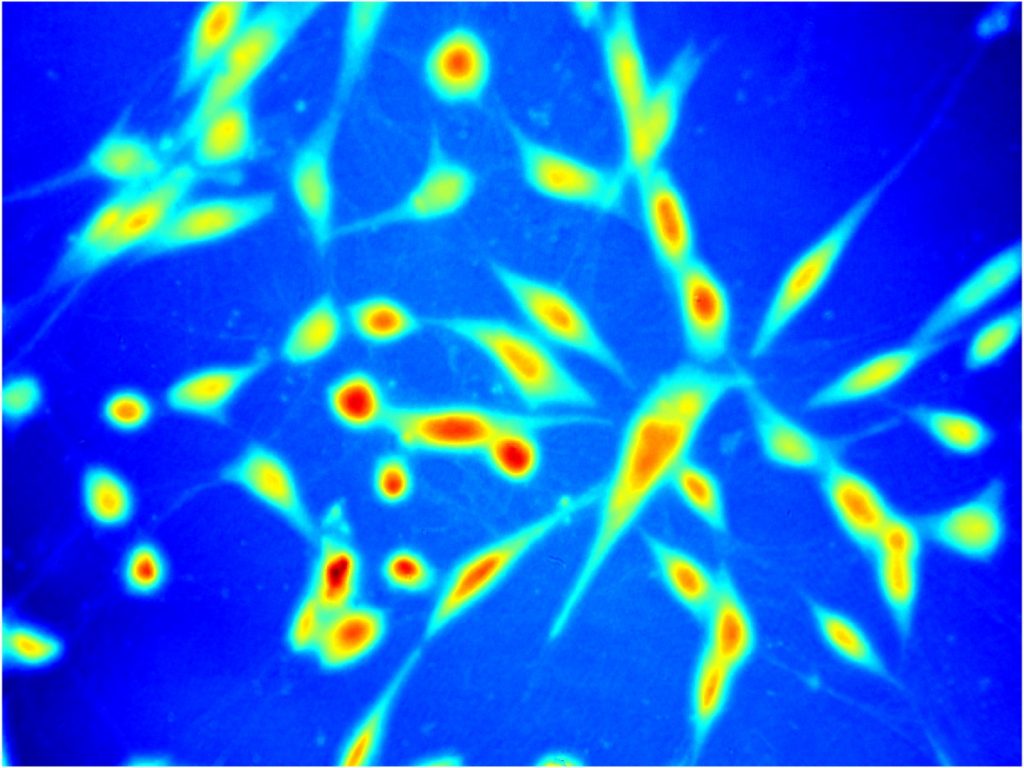
We started realization of a project “[GaboScope] Numerically enhanced lensless Gabor microscopy for high-throughput marker-free investigation of dynamic live biosamples“ funded (1,5 mln PLN) for 3 years (2021-2024) by National Science Center, Poland via SONATA programme. Principal investigator Maciej Trusiak and GaboScope Team are proud to cooperate with prof. Vicente Micó group (University of Valencia, Spain), prof. Chao Zuo group (Nanjing University of Science and Technology, China) and prof. Balpreet Ahluwalia group (The Arctic University of Norway, Tromso).
GaboScope Team: Julianna Winnik, PhD; Piotr Zdańkowski, PhD; Mikołaj Rogalski, MSc; Piotr Arcab, MSc and Maciej Trusiak, PhD
Interested in cooperation? Please contact us at maciej.trusiak@pw.edu.pl
OPUS: PHAICELL [Digital Holographic Microscopy]
We started realization of a project “[PHAICELL] Coherent quantitative phase microscopy: revisiting the basics and proposing novel numerical reconstruction methods with applications for advanced label-free bio-imaging” funded (2 mln PLN) for 4 years (2021-2025) by National Science Center, Poland via OPUS programme. Principal investigator Maciej Trusiak and PHAICELL Team are proud to cooperate with prof. Vicente Micó group (University of Valencia, Spain), prof. Chao Zuo group (Nanjing University of Science and Technology, China) and prof. Balpreet Ahluwalia group (The Arctic University of Norway, Tromso).
PHAICELL Team: Prof. Michał Józwik, Julianna Winnik, PhD; Piotr Zdańkowski, PhD; Maria Cywińska, MSc; Mikołaj Rogalski, MSc; Paweł Gocłowski, BSc; Jędrzej Szpygiel, BSc and Maciej Trusiak, PhD
Interested in cooperation? Please contact us at maciej.trusiak@pw.edu.pl
OPUS: [Digital Holographic Microscopy]
We started realization of a project “Numerically advanced phase and amplitude demodulation for optical interference microscopy and tomography“ funded (1.3 mln PLN) for 3 years (2018-2021) by National Science Center, Poland via OPUS programme. Principal investigator Maciej Trusiak.
PHAICELL Team: Prof. Michał Józwik, Julianna Winnik, PhD; Piotr Zdańkowski, PhD; Maria Cywińska, MSc; Mikołaj Rogalski, MSc; Paweł Gocłowski, BSc; Jędrzej Szpygiel, BSc and Maciej Trusiak, PhD
Interested in cooperation? Please contact us at maciej.trusiak@pw.edu.pl