Przejdź do treści

Check out our NEWS 2025

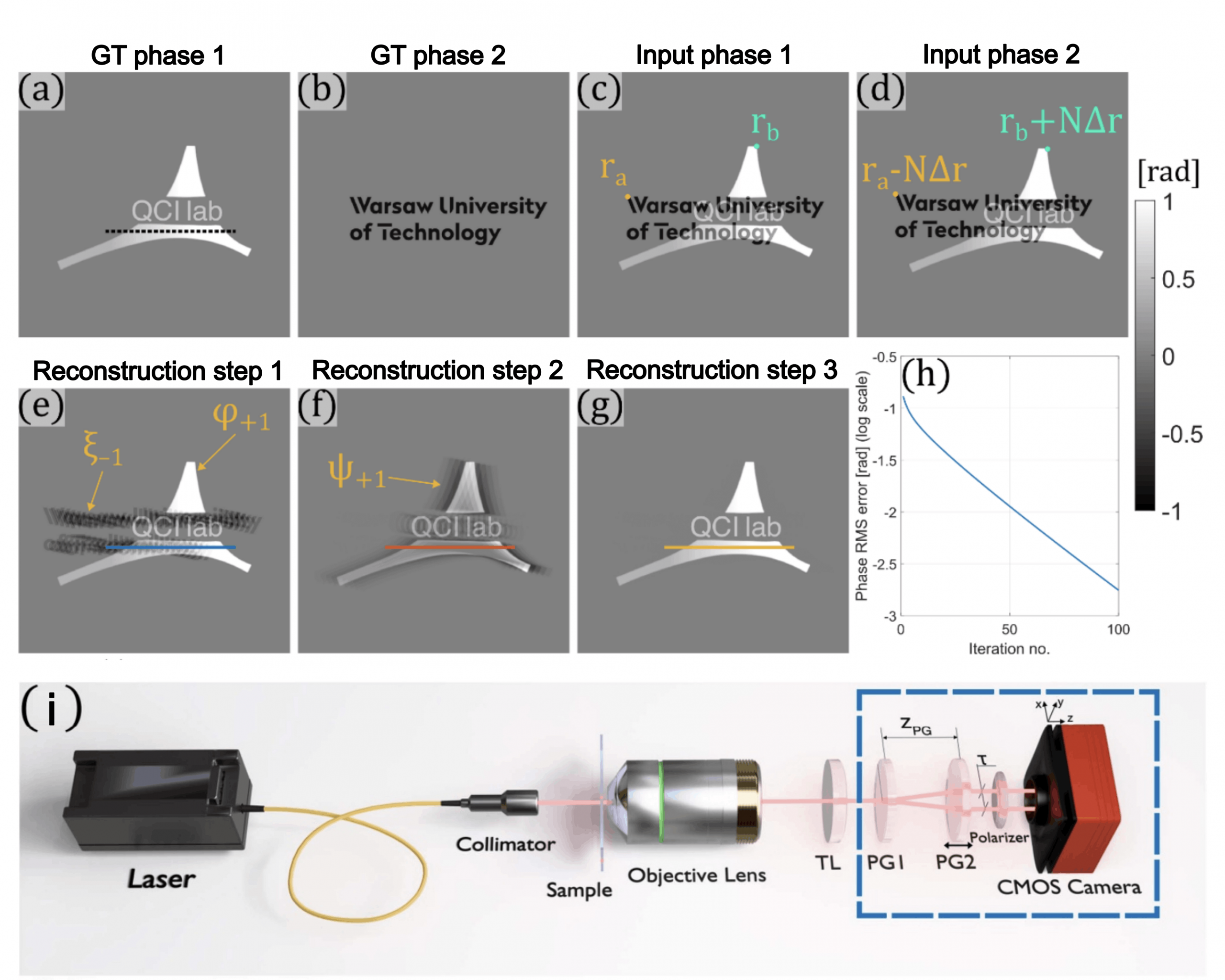
[December 2025] Check out our new publication in JPhys Photonics, included in Emerging Leaders 2025 collection: “Doubling the field of view and eliminating the replica overlap problem in common-path shearing quantitative phase imaging”.

🔍 Tackled problem
Common-path systems are significantly more robust to noise and vibrations than traditional dual-path configurations digital holographic microscopes. However, they have a major drawback: the two interfering object beams contribute to superimposed phase images in the reconstruction. This prevents imaging of dense, complex samples, restricting the method to sparse objects.

🛠️ Our solution
The key is our new MRR algorithm, which lies at the heart of the R2D-QPI method. It employs two or more separate measurements to analytically separate the overlapped phase images, effectively doubling the measured field of view.

🧑‍🔬Team
It was a result of fantastic cooperation led by QCI Lab: Mikołaj Rogalski, Piotr Zdankowski, Matyas Heto, Maciej Trusiak with Jolanta Mierzejewska, Małgorzata Lenarcik, Zhuoshi Li, Jiasong Sun and Chao Zuo.

Funding
Research was funded by the Project No. WPC3/2022/47/INTENCITY/2024 funded by the National Centre for Research and Development (NCBR) under the 3rd Polish-Chinese/Chinese-Polish Joint Research Call (2022). The research was conducted on devices cofounded by the Warsaw University of Technology within the Excellence Initiative: Research University (IDUB) programme. This work was supported by the National Natural Science Foundation of China (62227818, 62361136588, 62575139), National Key Research and Development Program of China (2024YFE0101300).

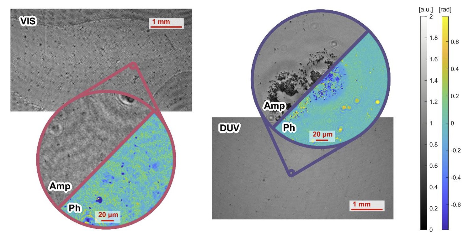
[December 2025] We are excited to share our new preprint: Low-dose Chemically Specific Bioimaging via Deep-UV Lensless Holographic Microscopy on a Standard Camera”.

🚀 We present a low-dose deep-UV lensless holographic microscopy platform that achieves large field-of-view DUV imaging (up to 116 mm²) with low illumination and simultaneous label-free phase and chemically specific amplitude contrast – all using standard CMOS sensors and no imaging optics. The system delivers submicron resolution and enables whole-slide, preparation-free bioimaging of cells, vesicles, and tissues, including label-free identification of lipid-rich hepatic stellate cells.

🧑‍🔬Team
Piotr Arcab, Mikolaj Rogalski, Karolina Niedziela, Anna Chwastowicz, Emilia Wdowiak, Julia Dudek, Julianna Winnik, Pawel Matryba, Jolanta Mierzejewska, Malgorzata Lenarcik, Ewa Stepien, Piotr Zdankowski, Grzegorz Szewczyk, Maciej Trusiak.

Funding
Funded by the European Union (ERC, NaNoLens, Project 101117392). Views and opinions expressed are however those of the author(s) only and do not necessarily reflect those of the European Union or the European Research Council Executive Agency (ERCEA). Neither the European Union nor the granting authority can be held responsible for them. The research was carried out using equipment co-funded by the Warsaw University of Technology within the Excellence Initiative: Research University (IDUB) program.

[December 2025] How do you calibrate a microscope without touching a single screw?
Our new method shows it’s possible — and fully automated!

We are pleased to announce our new publication in Optics and Lasers in Engineering:
“Two-step automated method for robust Fourier ptychographic microscopy LED misalignment calibration.” written by Piotr Arcab, Mikołaj Rogalski, Maciej Trusiak & Piotr Zdankowski

The whole article is available for free in full open-access: https://doi.org/10.1016/j.optlaseng.2025.109340

Fourier Ptychographic Microscopy (FPM) is a computational imaging technique that achieves both high resolution and a large field of view by synthesizing images captured under varying illumination angles.

Yet, its performance critically depends on accurate LED alignment — even slight positional or rotational errors can distort reconstructions.

Our study presents a fully automated, two-step calibration procedure that overcomes this limitation. The approach separately corrects translational and rotational LED array misalignments — without the need for specialized calibration targets, complex modeling, or iterative training.

By simplifying alignment while maintaining precision, this calibration strategy helps make FPM more accessible for digital pathology, biomedical imaging, and quantitative phase analysis — opening new opportunities for reproducible, high-quality optical imaging.

𝐖𝐡𝐚𝐭’𝐬 𝐧𝐞𝐱𝐭?
As said by the main author, Piotr Arcab:

Looking ahead, the next step is to integrate it seamlessly with motorized hardware and open-source software. Our long-term vision is to create a plug-and-play FPM system that requires no expert intervention and can be readily adopted in biomedical and clinical labs.

This work has been funded by the National Centre for Research and Development, Poland (LIDER14/0352/2023) in Lider XIV call and by the YOUNG PW grant financed by Warsaw University of Technology within the Excellence Initiative: Research University (IDUB) programme (Ministry of Science and Higher Education, Poland). The research was carried out on devices co-funded by the Warsaw University of Technology within the Excellence Initiative: Research University (IDUB) programme.

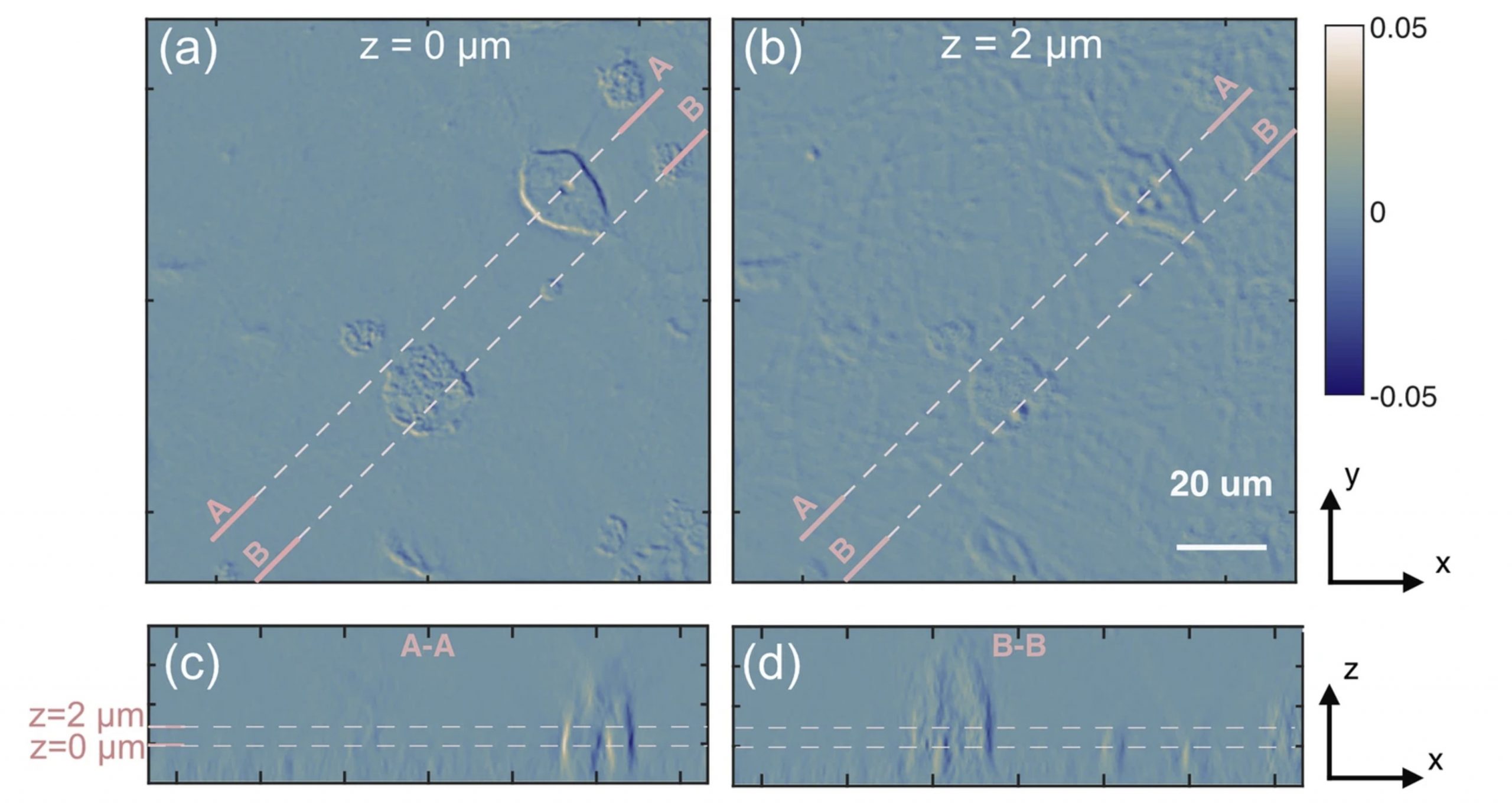
[November 2025] We’re excited to share our recent publication “Gradient Optical Diffraction Tomography (GODT): reconstruction of refractive-index gradients from phase-gradient measurements” in Communications Physics.

🔍 The novelty
Shearing and gradient-based phase measurements provide only phase derivatives, requiring fragile integration steps and often multiple z-scans to recover 3D refractive index. This limits robustness and quantitative accuracy. We introduce Gradient Optical Diffraction Tomography (GODT) — a rigorous tomographic framework that directly reconstructs the 3D refractive-index derivative from phase-gradient data. Validated with simulations, nano-printed phantoms, and fixed neural cells, GODT shows strong contrast and high sensitivity to refractive-index variations.

🧑‍🔬 Team
Developed through a great collaboration led by QCI Lab: Julianna Winnik, Piotr Zdańkowski, Maciej Trusiak, together with Marzena Stefaniuk, Azeem Ahmad, Balpreet Singh Ahluwalia, and Chao Zuo.

Funding
The work was funded by Sheng project: 2023/48/Q/ST7/00172, National Science Center, Poland
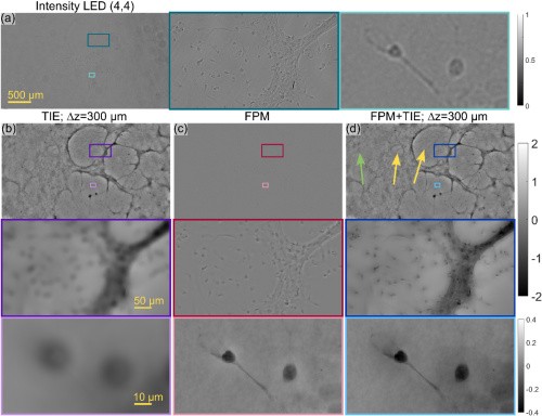
[November 2025] We’re happy to announce our new paper “Fourier ptychographic microscopy aided with transport of intensity equation for robust full phase spectrum reconstruction” in Optica Photonics Research.

🔍 Tackled problem
Fourier Ptychographic Microscopy (FPM) excels at high-resolution phase imaging, but low-frequency phase content is fundamentally difficult to recover under brightfield illumination.

🛠️ Our solution
We propose a hybrid FPM + Transport-of-Intensity (TIE) approach: a single additional on-axis defocused frame recovers missing low spatial frequencies via TIE, while standard FPM reconstructs high-frequency detail. The result is a quantitative, full-spectrum phase without overcomplicating the hardware.

👥 Team
Mikołaj Rogalski, Juan Martinez-Carranza, Bartosz Górski, Piotr Arcab, Michał Józwik, Piotr Zdańkowski, Magdalena Sobień, Marzena Stefaniuk, Shun Zhou, Chao Zuo, and Maciej Trusiak.

Funding
The work was supported by the National Centre for Research and Development (Project No. WPC3/2022/47/INTENCITY/2024) and Politechnika Warszawska (IDUB Young PW 504/04496/1143/45.010008)

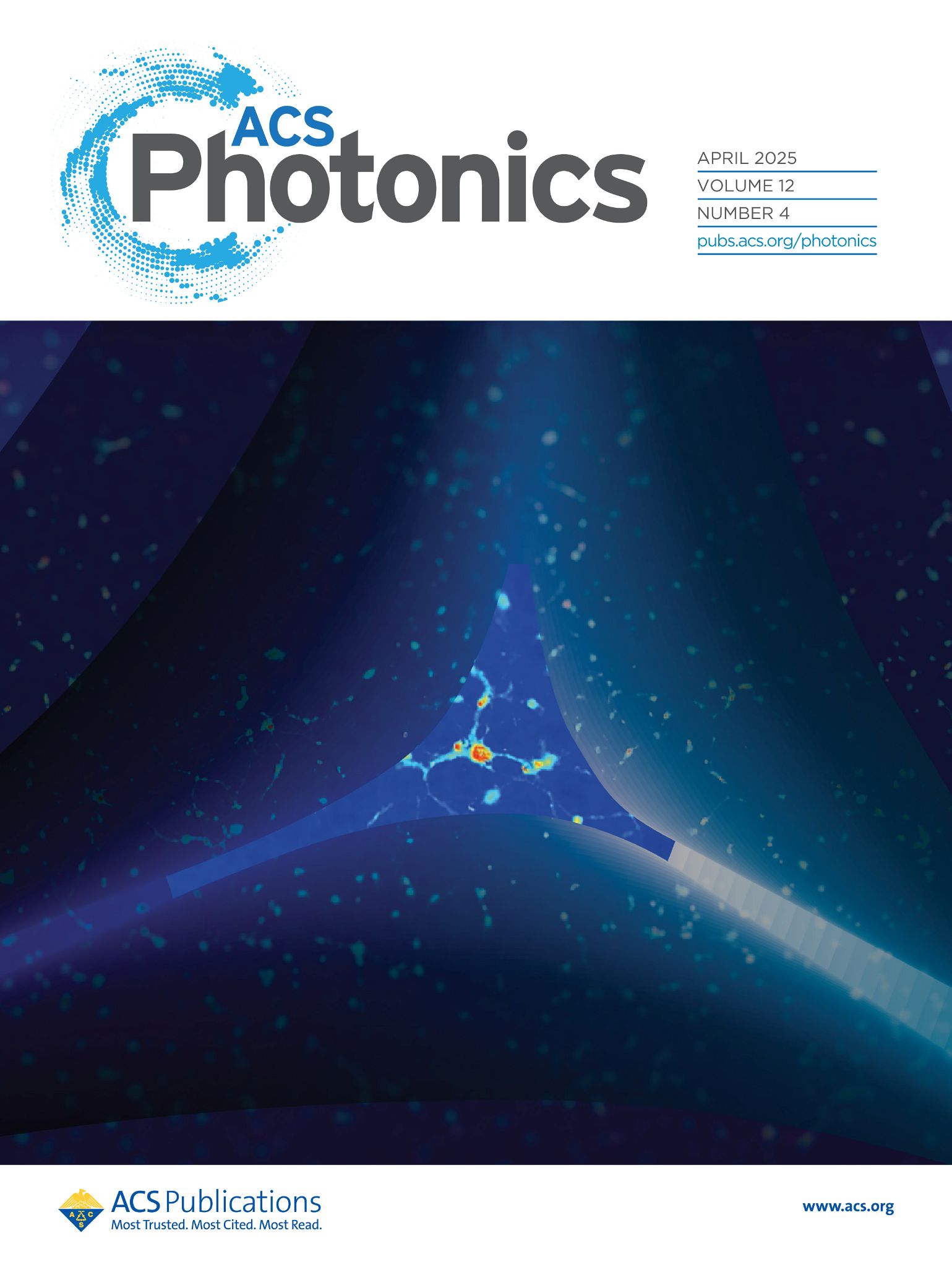
Alive cells with no stress. We are at ACS Photonics cover!

We demonstrated that you don’t need many photons to effectively observe living cells. Our researchers’ work has been recognized and featured on the cover of the prestigious journal ACS Photonics.

Our team has developed the iterative Gabor averaging (IGA) algorithm—a novel hybrid approach that integrates iterative phase retrieval with frame-averaging to suppress both twin-image artifacts and shot noise in multiframe digital in-line holographic microscopy (DIHM).

Thanks to this algorithm, we can image samples at even lower illumination intensities than before. That’s crucial when using our microscope to study live cell colonies—because even low light levels can stimulate or damage them, skewing the results

emphasizes Dr. Eng. Mikołaj Rogalski, lead author of the publication.

Our scientists are not slowing down. As Dr. Rogalski reveals:

Our next goals are to further improve noise reduction under low-illumination imaging conditions. We plan to apply the algorithm to real biological studies to minimize phototoxicity’s impact on cell-migration analysis.

🧾 Read more in our paper:
Hybrid Iterating-Averaging Low Photon Budget Gabor Holographic Microscopy
Autors: Mikolaj Rogalski, Piotr Arcab, Emilia Wdowiak, José Ángel Picazo-Bueno, Vicente Micó, Michał Józwik, Maciej Trusiak
https://doi.org/10.1021/acsphotonics.4c01863

💎 The research was funded by:
1) the European Union (European Research Council (ERC), NaNoLens, Project 101117392);
2) the Warsaw University of Technology within the Excellence Initiative: Research University (IDUB) program
3) the Grant PID2020-120056 GB-C21 funded by MCIN/AEI/10.13039/501100011033. José Ángel Picazo Bueno (Universitat de València) is supported by the Spanish grant “Margarita Salas” (ref MS21-100), proposed by the Ministerio de Universidades (UP2021-044) funded by the European Union, NextGenerationEU.
[ July  2025 ] We go to Ångström-resolution on our seminar 🔬

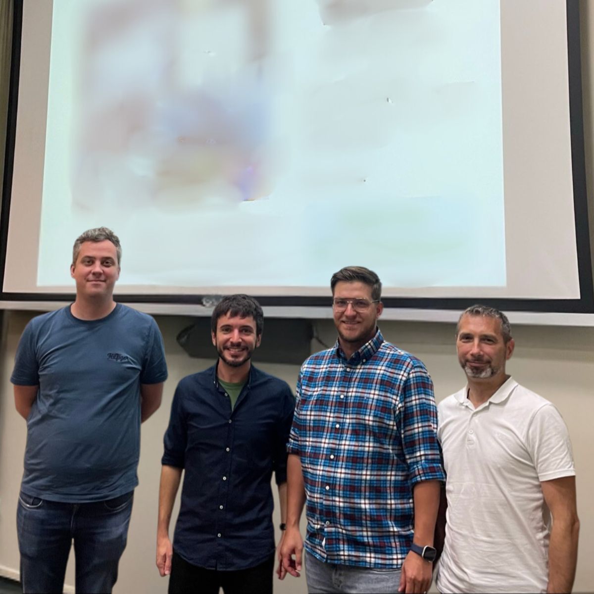
Dr Luciano A. Masullo shared his results in Ångström-resolution fluorescence microscopy, giving us a high dose of inspiration thanks to him.

He is a Project Leader at Max Planck Institute of Biochemistry and a graduate of the Universidad de Buenos Aires. He is an expert in super-resolution microscopy.

Dr Masullo has a remarkable talent to describing very complex and groundbreaking scientific topics in a precise and easy-to-understand way.

The idea that every human cell is covered by sugars (carbohydrates) will stay with us for a long time 💡

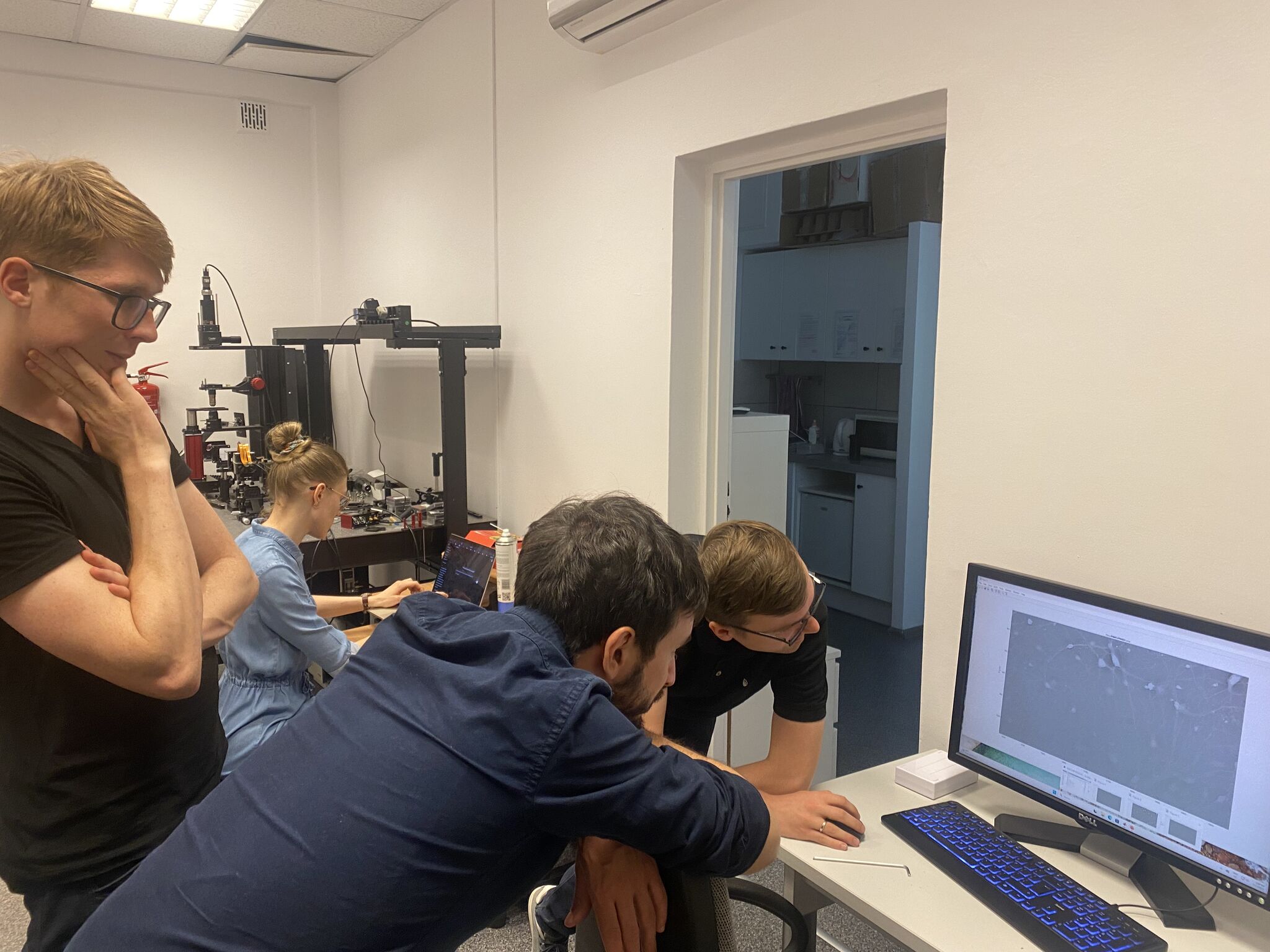
After his speech, we showed him around our lab and had great discussions about microscopy🦠

We can’t wait to meet on the next occasion!
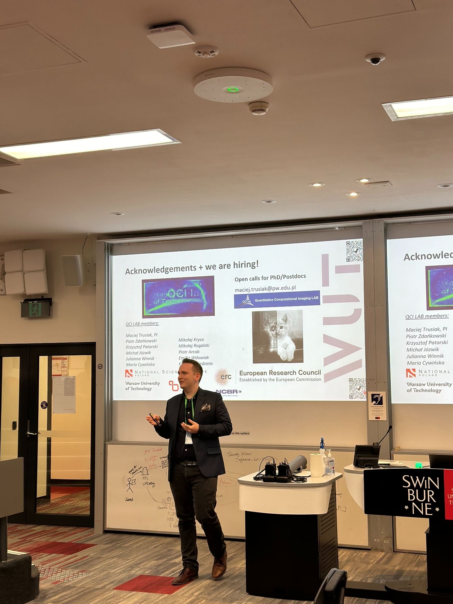
[ June  2025 ] Sometimes science requires very long journeys…
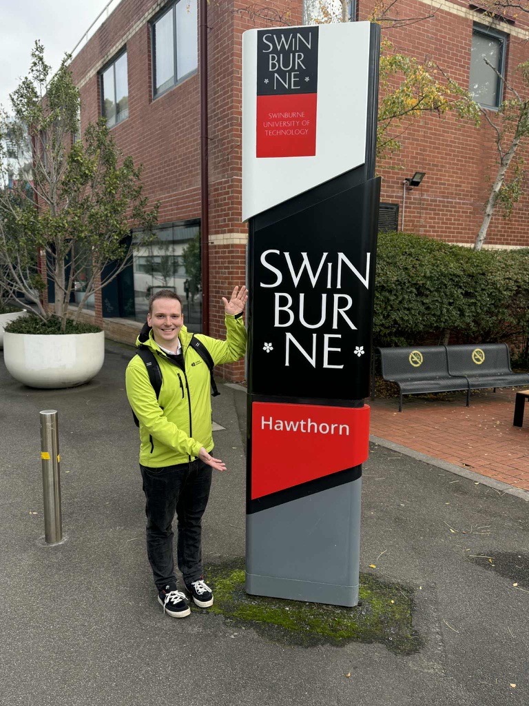
That’s why our science voyager visited Australia!

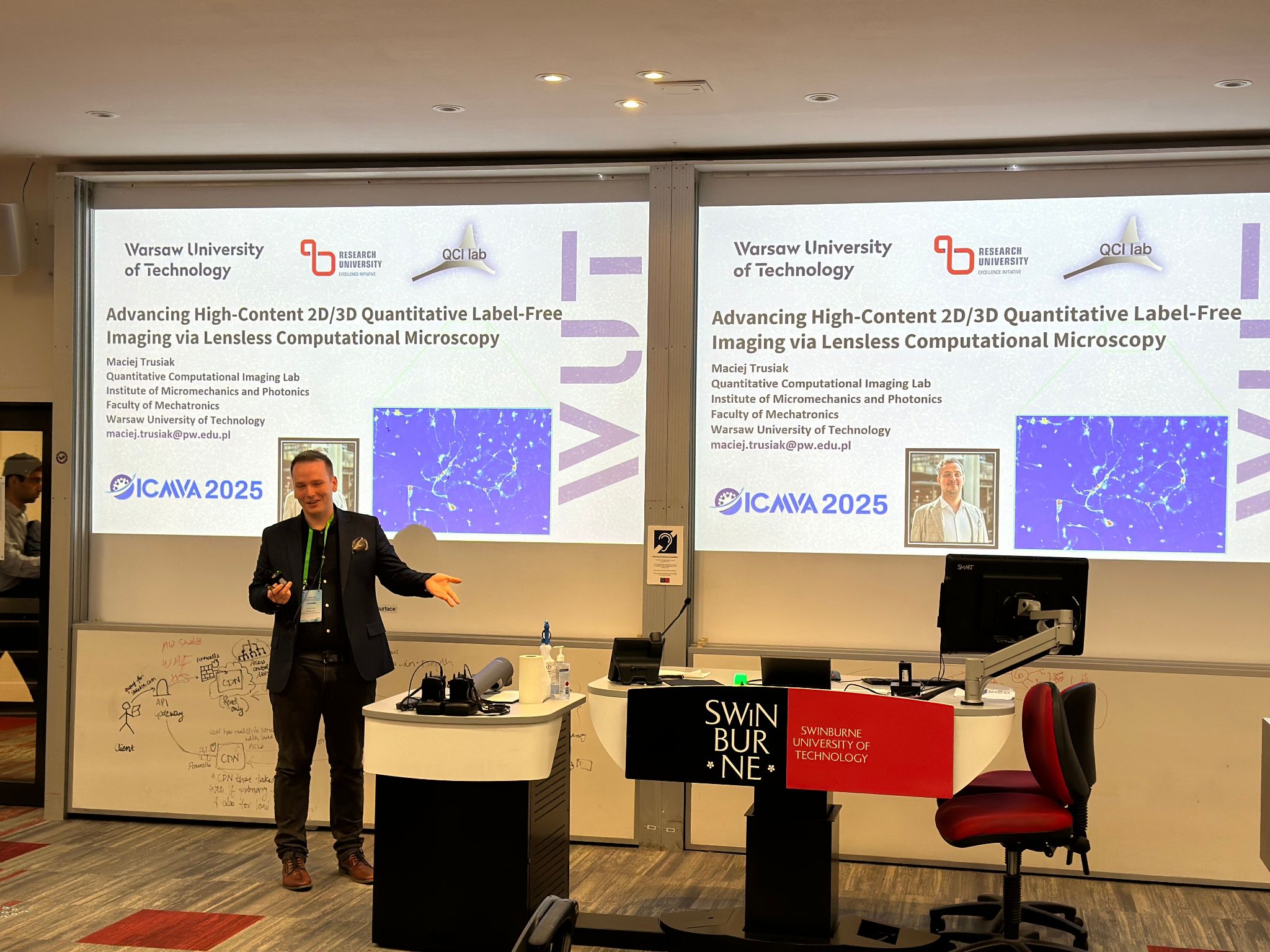
Piotr Arcab travelled over 15,000 km from Warsaw University of Technology to Melbourne, Australia to spread the word about Lensless Microscopy 🔬

He visited the prestigious 8th International Conference on Machine Vision and Applications. Piotr showed our results in the presentation:
🔹 Advancing High-Content 2D/3D Quantitative Label-Free Imaging via Lensless Computational Microscopy 🔹

Huge thanks to Vijayakumar Anand for invitation! We can’t wait for the next opportunity to collaborate.

The research was funded thanks European Research Council (ERC) Starting Grant: NaNoLens project [Lensless microscopy]

Piotr’s journey was funded thanks to IDUB Mobility grant of Warsaw University of Technology.

[ May  2025 ] 🎉 Congratulations to Our Outstanding PhD Students!

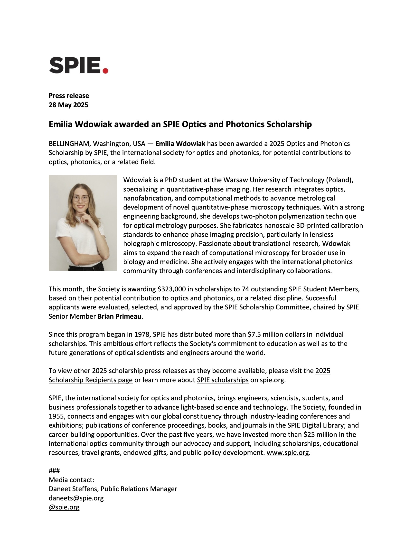
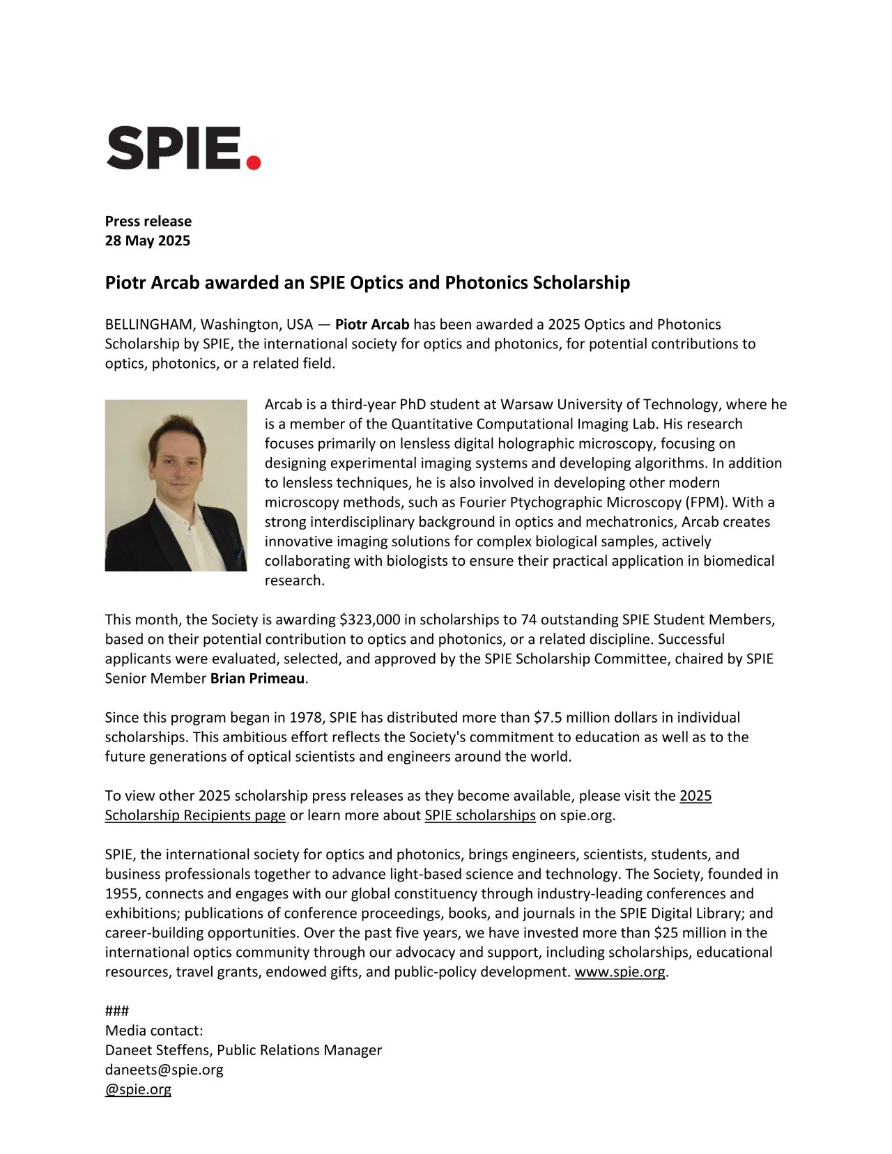
We are thrilled to announce that our PhD students, Emilia Wdowiak and Piotr Arcab, have been awarded the prestigious 2025 Optics and Photonics Scholarship from SPIE, the international society for optics and photonics.

This year, only 74 students worldwide received this recognition — and Emilia and Piotr are the only two recipients from Poland. This competitive scholarship highlights their exceptional academic achievements, dedication to research, and significant contributions to the field of optics and photonics.

Their work continues to inspire and advance our scientific community. 👏
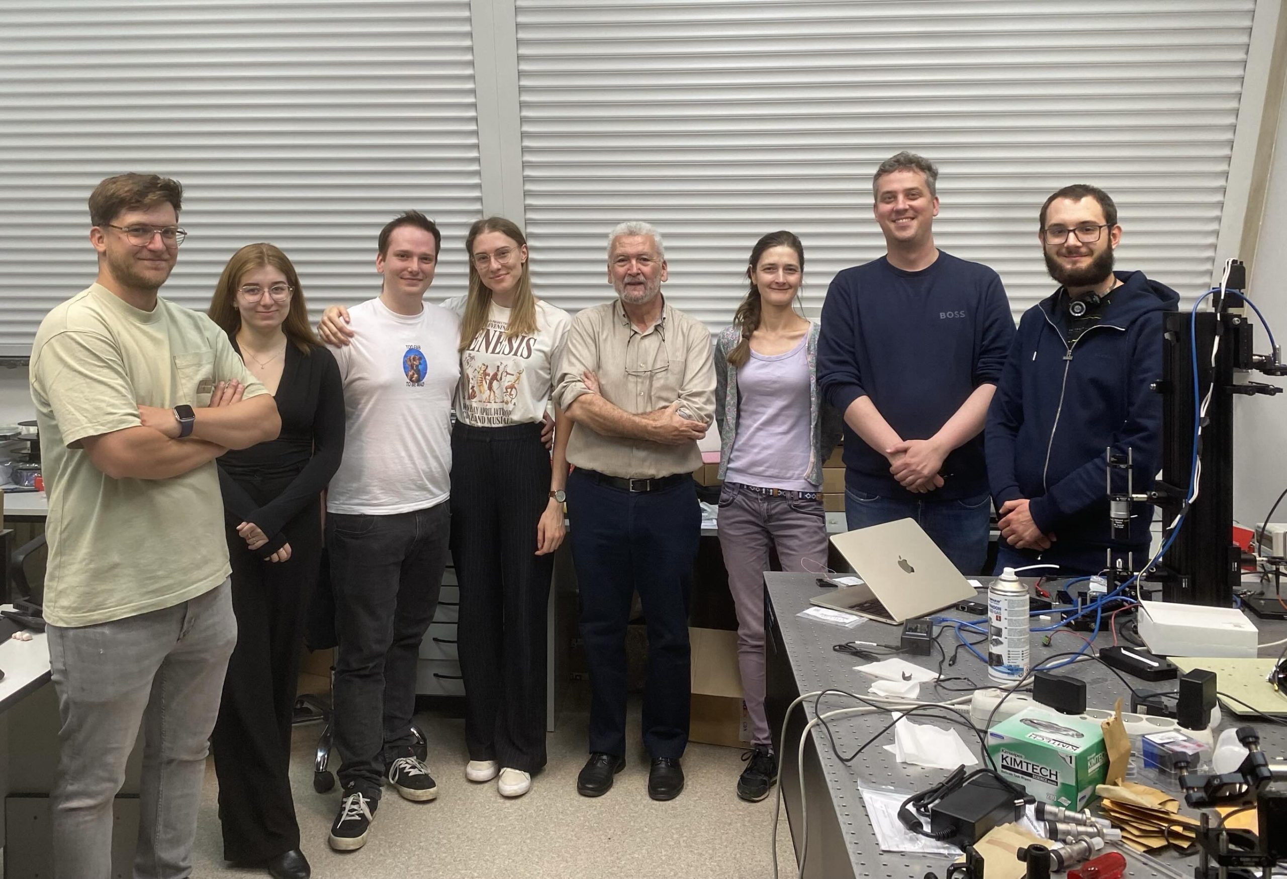
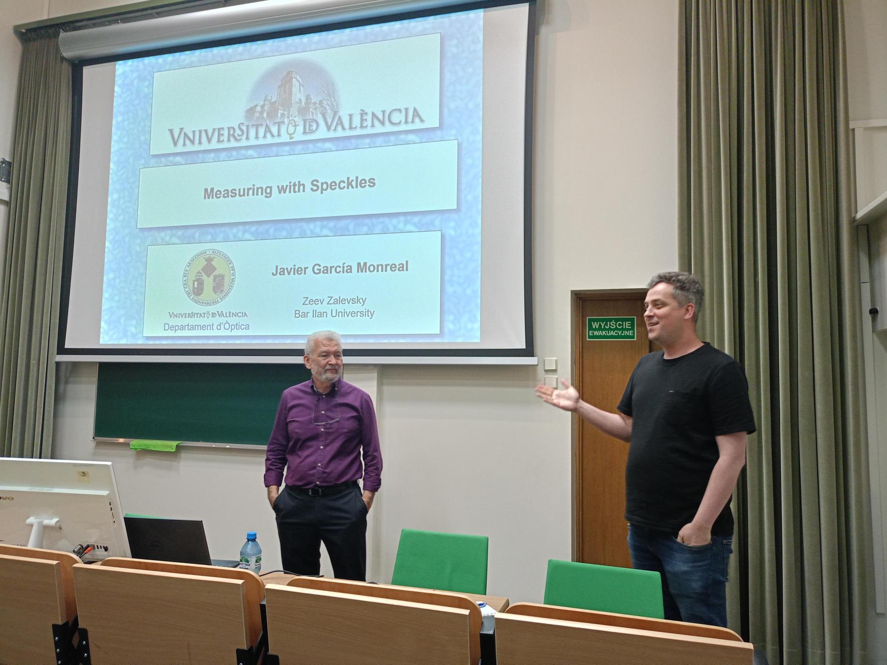
[20 – 21 May  2025] We We were delighted to host Prof. Javier García, a distinguished researcher from the University of Valencia and member of the Photonics and Optical Engineering Group.

During his visit, Prof. García was given a guided tour of our research facilities, where he had the opportunity to engage with our teams and learn about our latest work in optical imaging and photonics. His visit sparked lively discussions and fostered new ideas for potential collaborations.

One of the highlights of his stay was his captivating lecture on imaging techniques using speckles, where he shared innovative approaches and recent advancements in the field. The talk attracted a broad audience from across our colleagues.

We thank Prof. García for his visit, for sharing his insights, and for inspiring our community with his work. We look forward to future exchanges and collaborations!

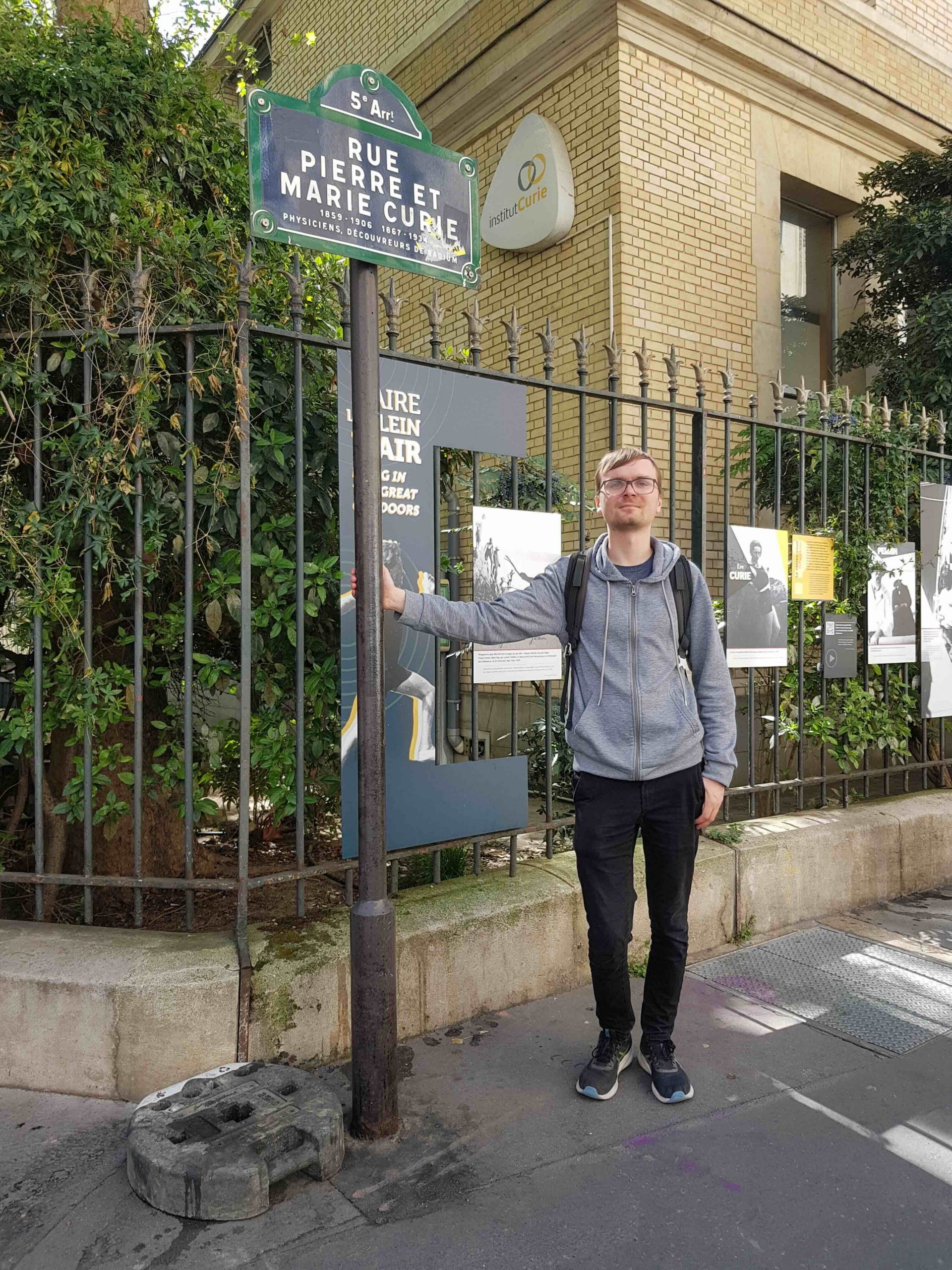
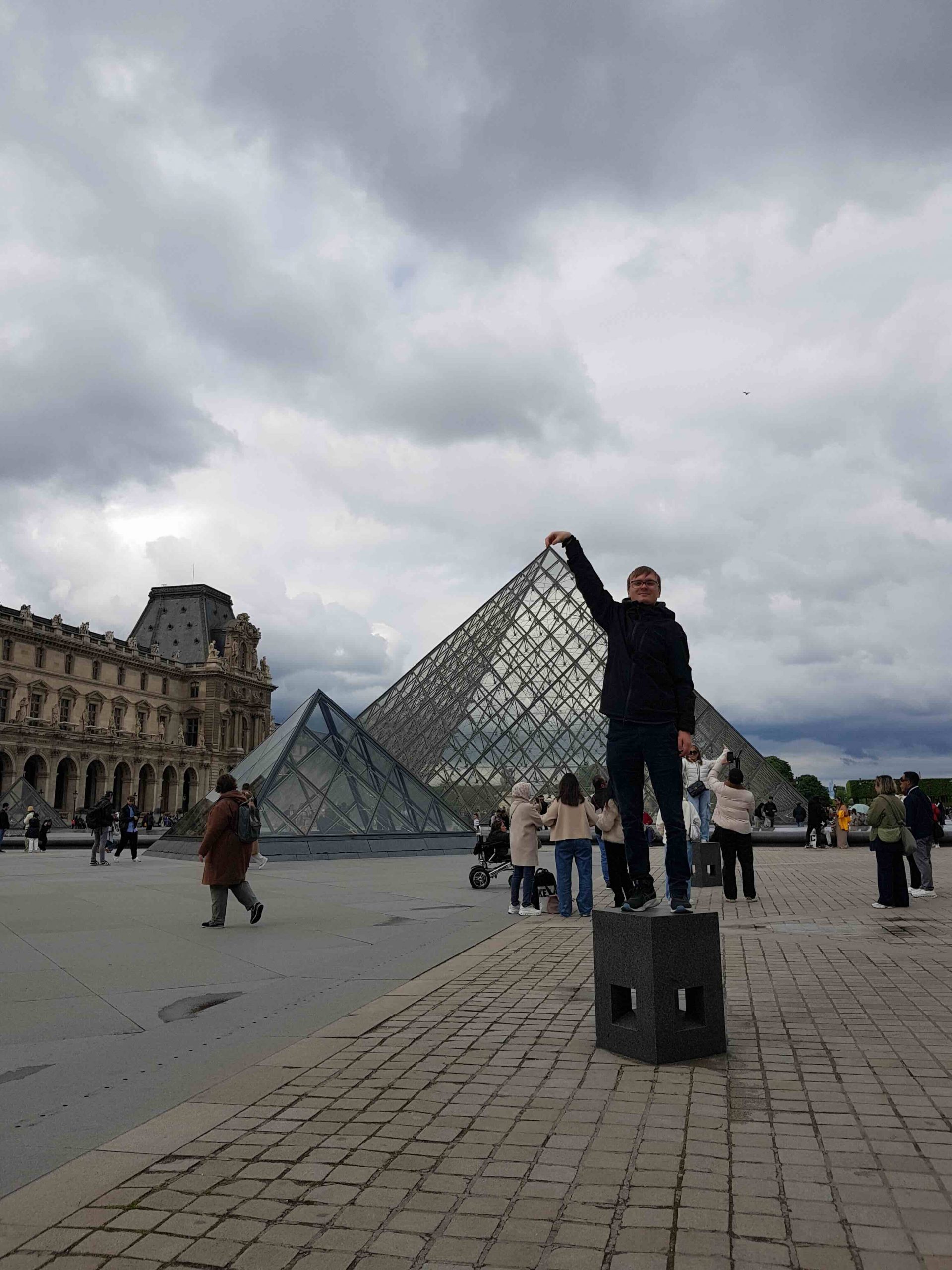
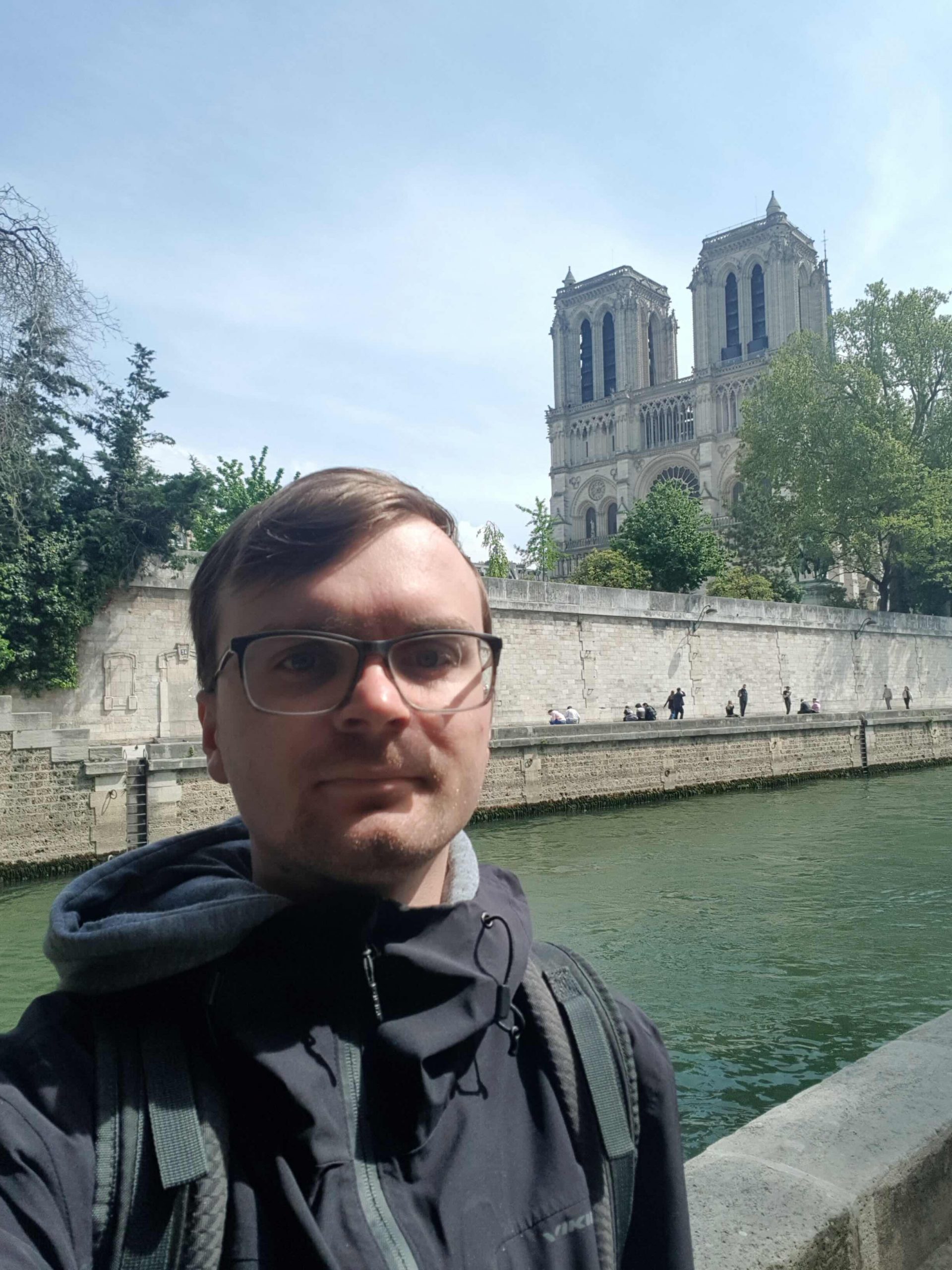
[February – April  2025] We are excited to share that our beloved PhD Mikołaj Rogalski recently completed a three-month research stay at the prestigious Kastler Brossel Laboratory in Paris, within the Complex Media Optics group led by Prof. Sylvain Gigan and Dr. Hilton Barbosa de Aguiar.

During his visit, Mikołaj focused on developing advanced phase imaging techniques for samples embedded between strongly scattering media. His work aimed to improve the precision and robustness of phase retrieval in complex optical environments – an important step forward for applications in biomedical imaging and optical diagnostics.

Working closely with Complex Media Optics team, Mikołaj benefited from the group’s expertise in imaging through complex media and cutting-edge experimental techniques. The interdisciplinary and collaborative environment at the Kastler Brossel Laboratory greatly enriched his research and opened new directions for future exploration in the field of optical imaging.

His stay in Paris was supported by the Mobility PW travel grant, awarded as part of the XIII edition of the programme within IDUB initiative and by the ERC Starting Grant: NaNoLens project.

We are proud of Mikołaj’s accomplishments and grateful to Professor Sylvain, Dr. Hilton and the Complex Media Imaging group for their warm welcome and fruitful collaboration. We look forward to strengthening the ties between our institutions and to future joint research endeavors.

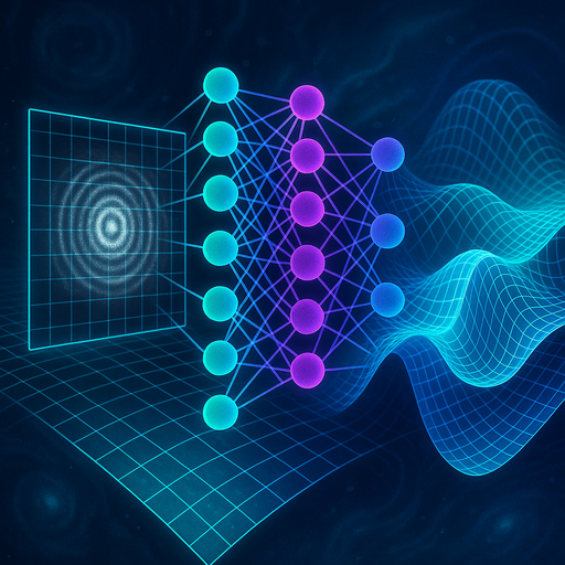
AI-generated artistic vision of DeepQuadrature

[April  2025] DeepQuadrature: from single-shot to accurate laboratory measurements

Researchers from the QCI Lab at the Faculty of Mechatronics, Warsaw University of Technology, in collaboration with UiT The Arctic University of Norway and the University of Münster, have developed an innovative AI-powered tool that could make the analysis of optical interferograms and holograms easier. Their findings were published in the prestigious Journal of Physics: Photonics. 

M. Cywińska, M. Józwik, K. Patorski, A. Ahmad, B. Ahluwalia, B. Kemper, M. Trusiak,DeepQuadrature: universal convolutional neural network enlarging space–bandwidth product in single-shot fringe pattern optical metrology, J. Phys. Photonics vol. 7 (2025)

DOI 10.1088/2515-7647/adc219https://iopscience.iop.org/article/10.1088/2515-7647/adc219

DeepQuadrature is a deep learning model capable of accurately estimating the so-called quadrature function—an image phase-shifted by π/2 relative to the input fringe pattern. This enables precise reconstruction of phase distributions using only a single measurement frame, significantly simplifying and accelerating optical measurements, while eliminating the need for traditional multi-frame methods. 

What’s the Challenge? 

Fringe-pattern-based measurement techniques—such as holographic microscopy or interferometry—are widely used in biology, optics, and materials engineering. However, their accuracy is fundamentally limited by the space-bandwidth product (SBP), which defines how much and how detailed information can be extracted from an image. 

DeepQuadrature boosts SBP purely numerically, without requiring any changes to the optical setup (fig. 1). Trained on synthetic datasets with diverse fringe geometries and frequencies, the model can adaptively analyze both technical and biological objects—from metal surface microstructures to HeLa cell imaging. In experimental tests, QCI Lab researchers achieved accuracy comparable to state-of-the-art multi-frame approaches, while maintaining the simplicity of single-frame data acquisition. 

 
Fig. 1. Principle of operation of DeepQuadrature. 

Multi-frame techniques require costly hardware capable of acquiring phase-shifted image sequences. DeepQuadrature offers a software-based alternative to expensive hardware components.  
As the lead author, Maria Cywińska, PhD, explains,  

The benefits go far beyond cost savings—single-shot measurements are faster and easier, making them suitable for broader applications, including clinical diagnostics. This paves the way for bringing measurements out of the lab.

What’s Next? 

Our team trained DeepQuadrature as a general-purpose model, capable of handling a wide range of optical metrology tasks.  
Looking ahead, Maria Cywińska, PhD, concludes:  

In future work, we aim to specialize the network by training it for specific experimental applications, such as imaging a particular type of biological sample. This will allow us to directly compare the performance of generalist vs. task-specific models. 

Get the Model 

The DeepQuadrature model and the datasets used for training and validation are freely available on GitHub: github.com/MariaSi1/DeepQuadrature

Contact Us 

We are happy to collaborate or answer questions related to this research.
Feel free to reach out:
maria.cywinska@pw.edu.pl  

Team & Funding 

This work was carried out within the QCI Lab team (Maria Cywińska, PhD, Prof. Michał Jóźwik, Prof. Maciej Trusiak) in collaboration with the Arctic University of Norway in Tromsø (Azeem Ahmad, PhD, Prof. Balpreet Ahluwalia) and the University of Münster (Prof. Bjorn Kemper).
This work has been partially funded by the National Science Center Poland (PRELUDIUM 2021/41/N/ST7/04057 and OPUS 2020/37/B/ST7/03629) and the Ministry of Science and Higher Education (Polish Metrology PM/SP/0008/2021/1).
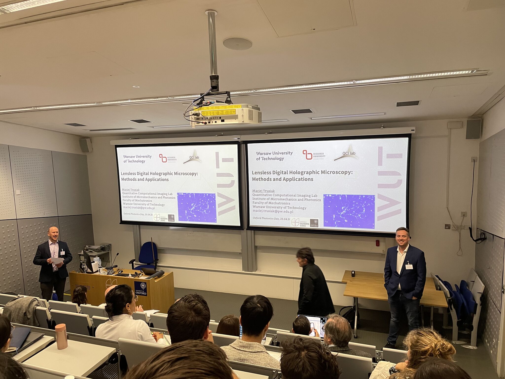
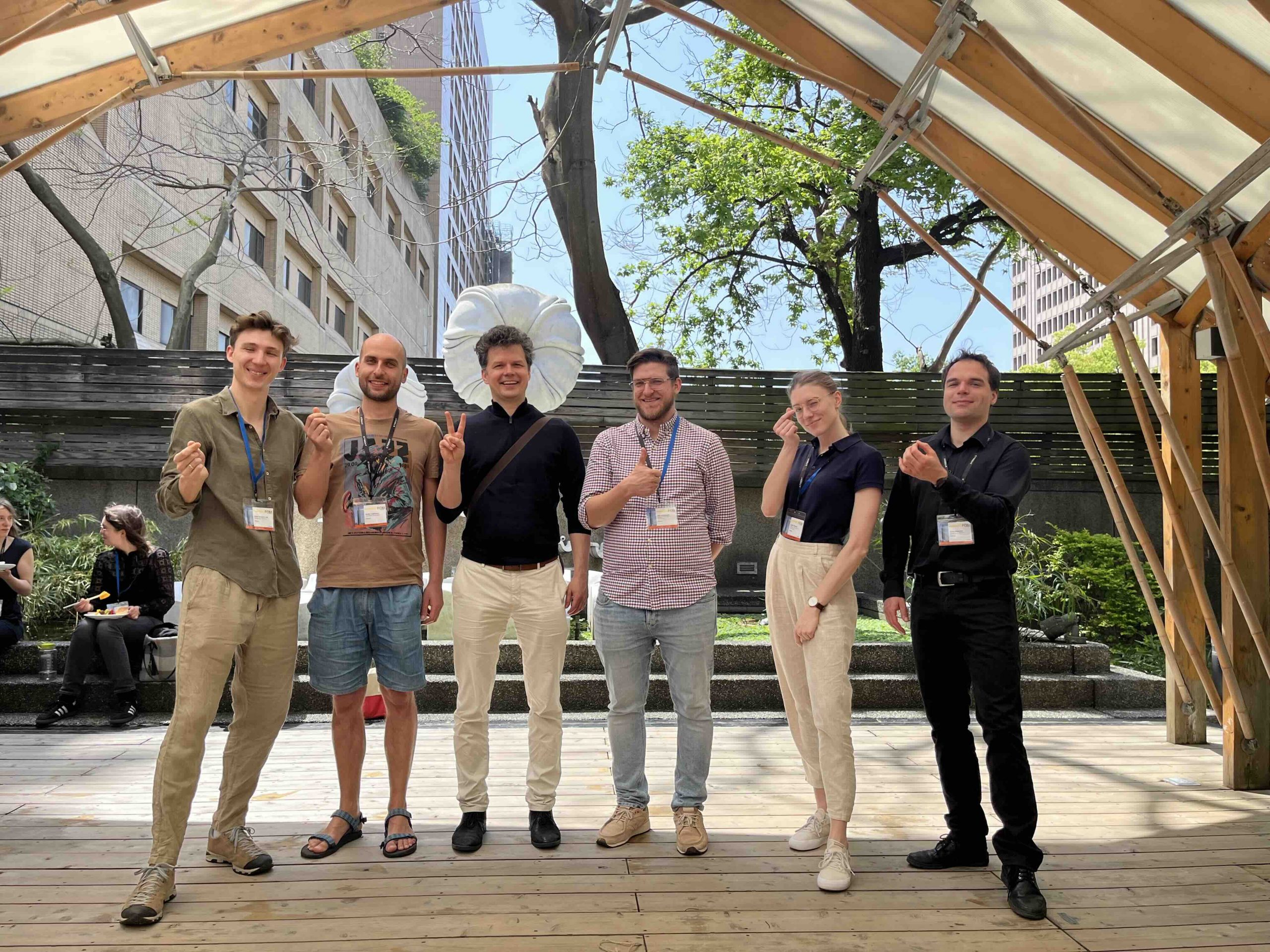
[25  April  2025]  Maciej Trusiak and Piotr Zdańkowski took part in Oxford Photonics Day 2025 taking place in the Department of Engineering Science at the University of Oxford.

Maciej delivered an invited talk on “Lensless Digital Holographic Microscopy: Methods and Applications”.

Piotr presented some of his novel research results during the poster session.


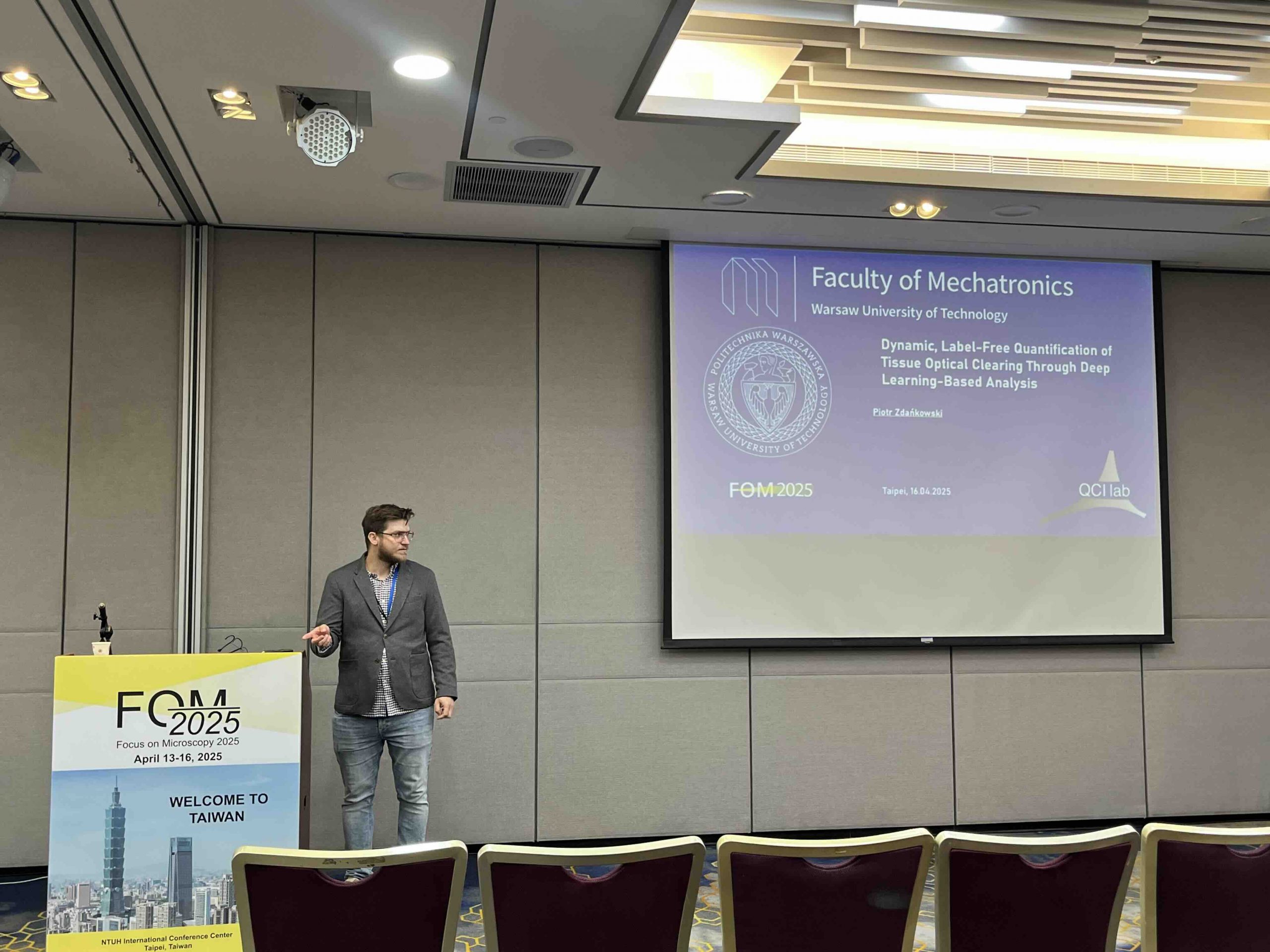
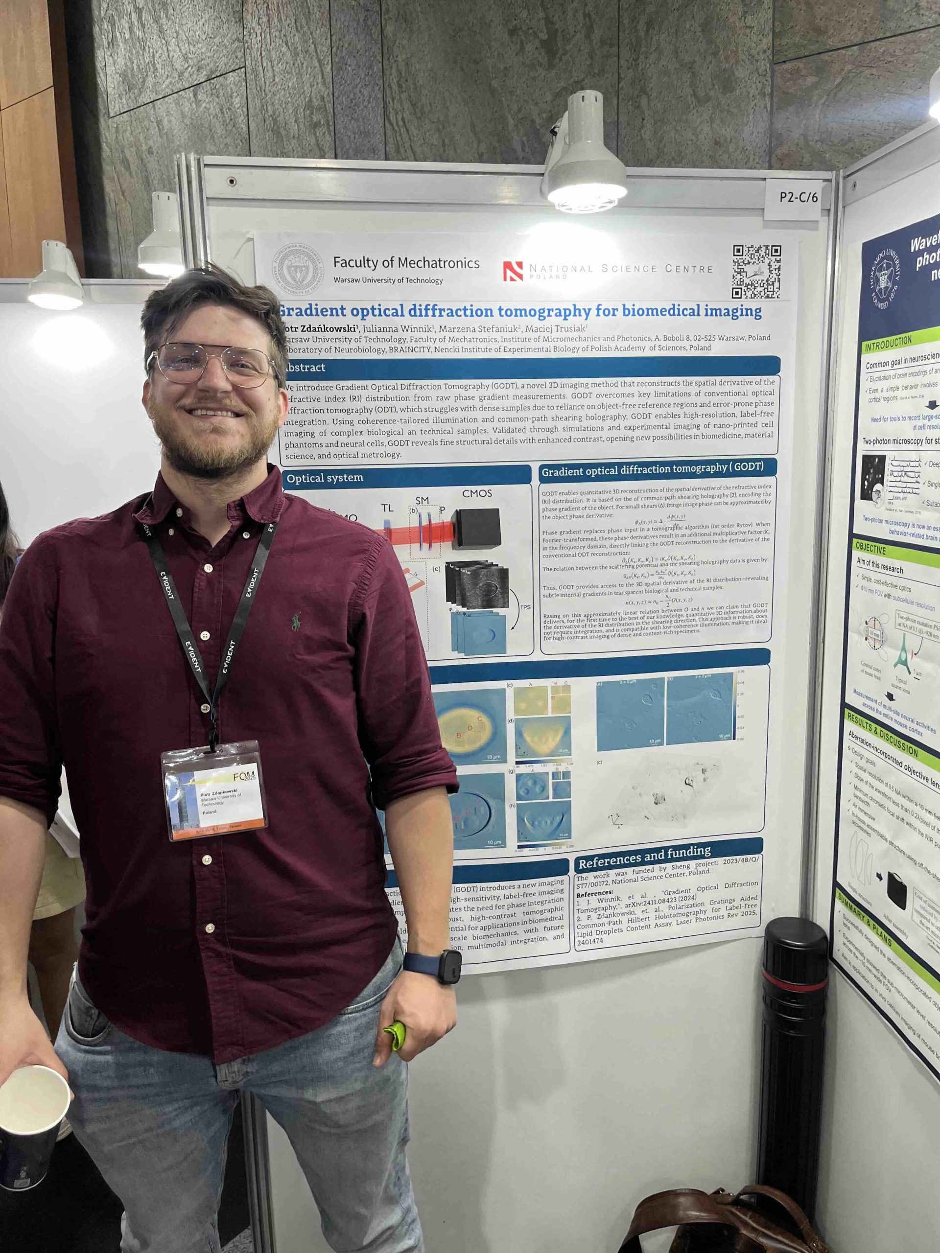
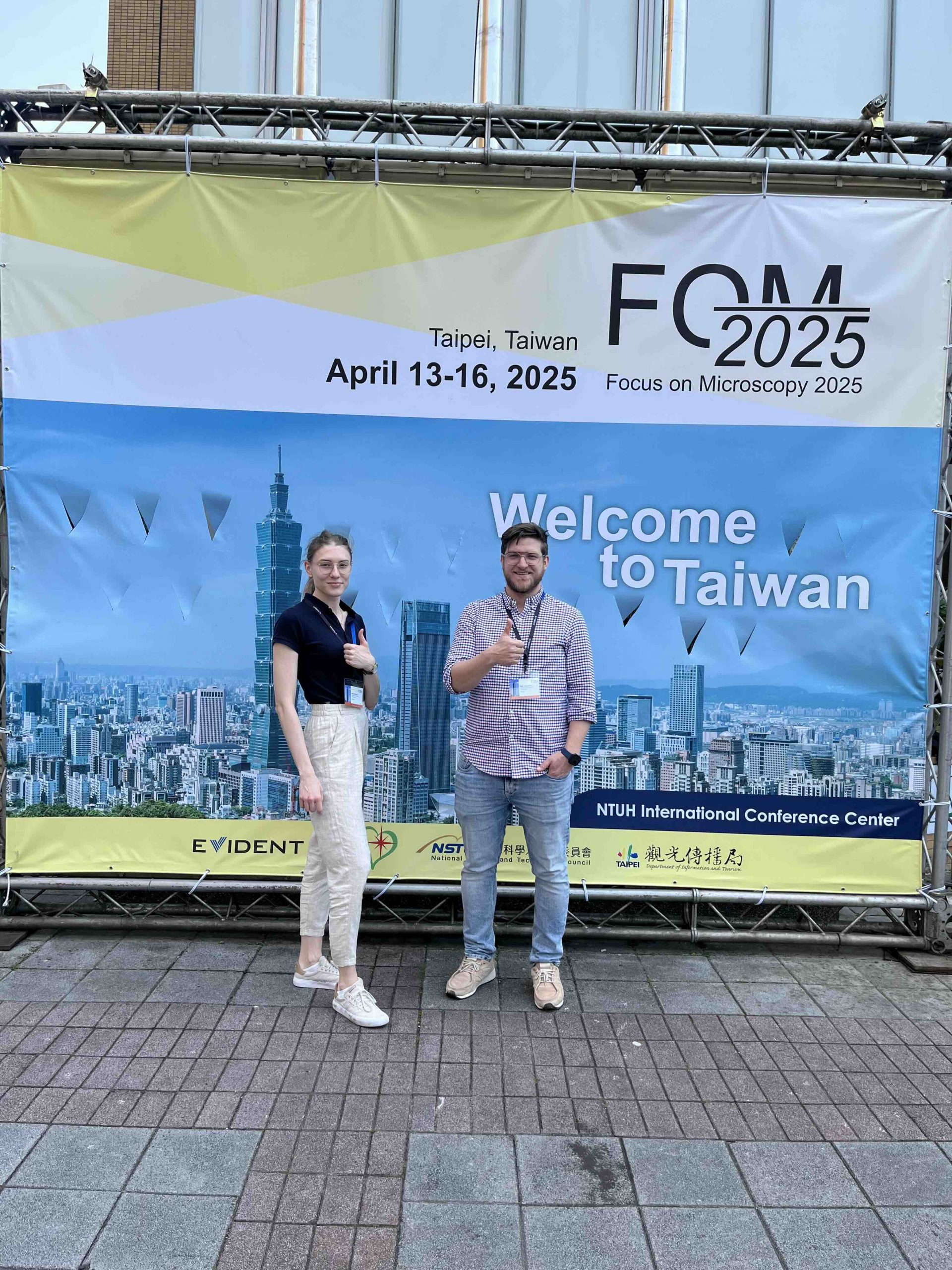
[13 – 16  April  2025]  Piotr Zdańkowski and Emilia Wdowiak attended Focus on Microscopy 2025 conference (FOM 2025) taking place in Taipei, Taiwan.

Piotr gave a talk on “Dynamic, Label-Free Quantification of Tissue Optical Clearing Through Deep Learning-Based Analysis” and presented a poster on “Gradient optical diffraction tomography for biomedical imaging”.

Emilia delivered a talk concerning “In-line holographic microscopy with liquid crystal speckle reducers”.

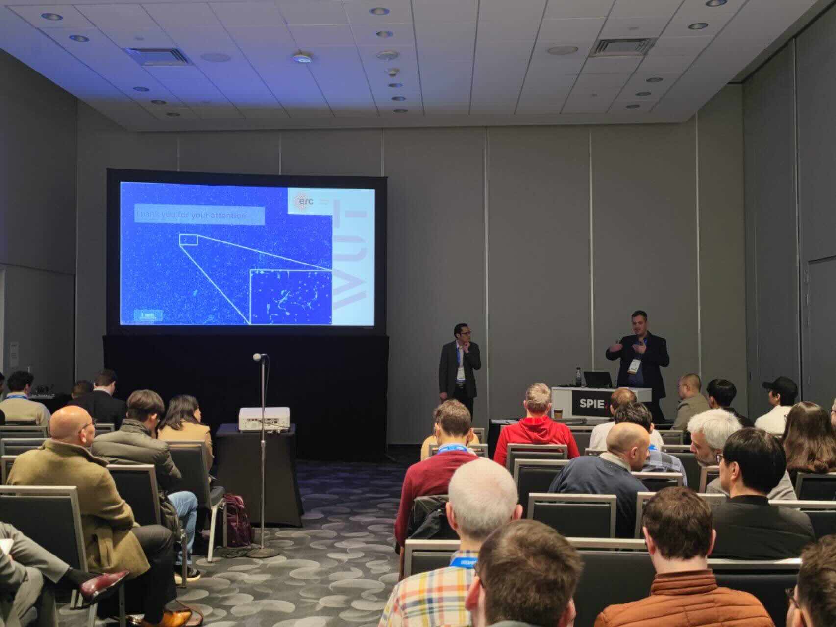
[25 – 30  January  2025] Maciej Trusiak represented QCI lab at Photonics West 2025 in San Francisco, CA USA.

He contributed to the Quantitative Phase Imaging XI conference and had a pleasure to give an invited talk about “Large field-of-view lensless digital holographic microscopy for cell and tissue imaging”. He presented some of the latest works that our group is working on. 

Photonics West is always a great place to meet world-class scientists and learn about the novelties in the field.

[January  2025] Julianna Winnik was nominated as a Rising Star in Optics 2024 – a programme designed to honour outstanding early-career researchers who are making significant contributions in optics.

CONGRATULATIONS! Very well-deserved 🥳


[3 January  2025]   Prof. Maciej Trusiak took a part in the 1st International Conference on Emerging Trends in Optical Technologies ETOT 2025.

He had a chance to give a keynote speech concerning the advancements in holographic imaging methods.


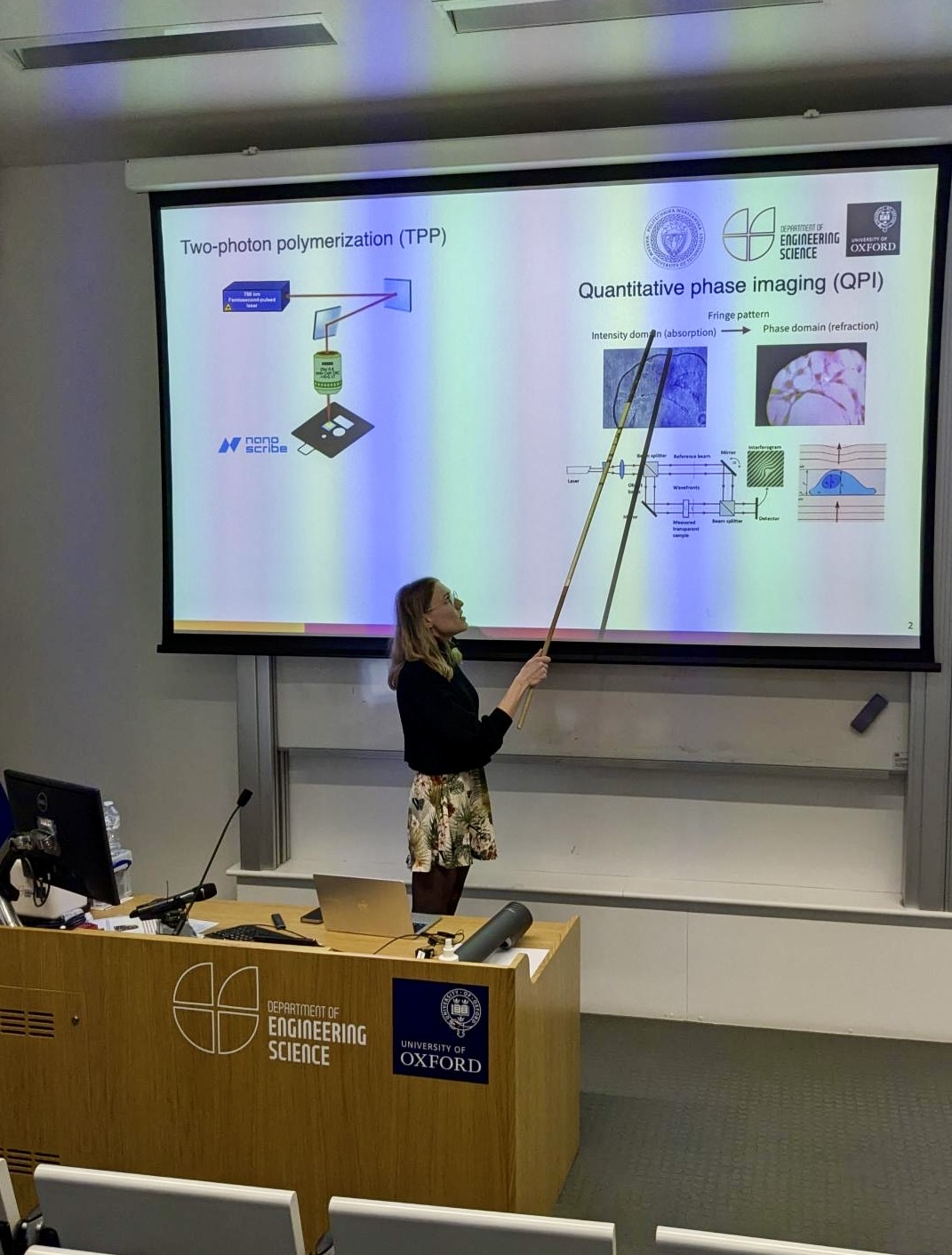
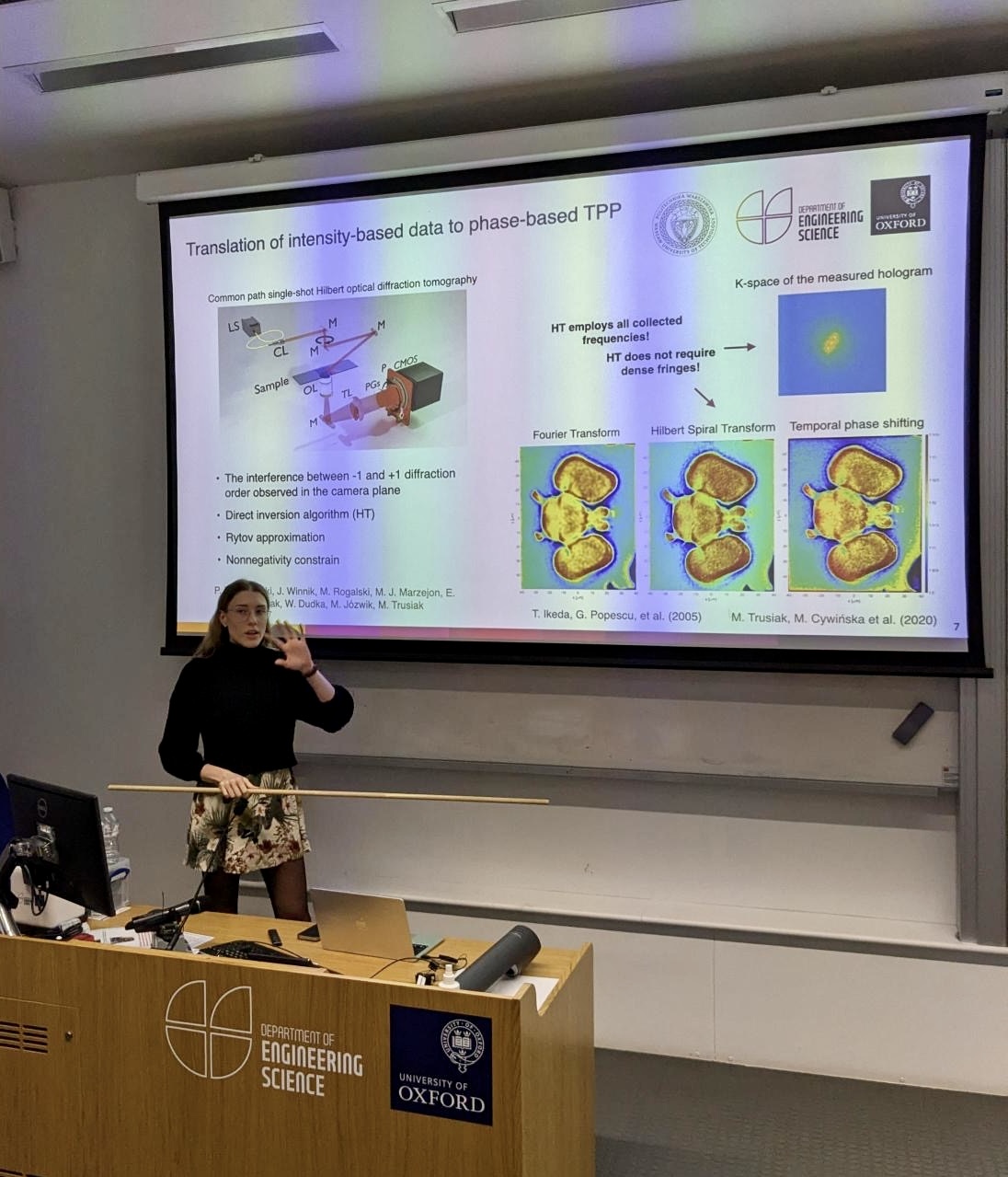
[September – December  2024] We are thrilled to share that our PhD student, Emilia Wdowiak, spent three months collaborating with Professor Martin Booth‘s group at the University of Oxford, Department of Engineering Science.

During her stay, Emilia developed a cutting-edge quantitative phase imaging method for laser-written liquid crystal cells. Advantaging the exceptional resources of the University of Oxford, she deepened her knowledge of innovative liquid crystal technologies and their groundbreaking applications. She also had the opportunity to contribute to ongoing research projects, including liquid crystal two-photon polymerization and the group’s unique speckle reduction techniques.

Her visit was supported by the Mobility PW travel grant, which she earned as part of the XIII edition of the programme. This initiative is part of the “Research Initiative – Research University” SEED and PROM PW 2 projects.

Emilia’s time in Oxford provided invaluable insights for her PhD research and greatly contributed to her scientific growth. We are thankful to the University of Oxford and Professor Booth’s group for their collaboration, and we look forward to the future cooperation between Oxford and Warsaw!